
3.4 Conception and Prenatal Development
When we imagine pregnancy, often think of it in terms of time: weeks, months, trimesters. Pregnancy is a dynamic process, with both mother and fetus changing almost daily. Beginning with conception, prenatal development is organized by stages: the germinal period, embryonic period and fetal development period. To help our understanding of what happens during conception, let’s first take a closer look at the building blocks of our DNA.
3.4.1 Stages of Prenatal Development
Conception has occurred, and the fertilized egg has been implanted in the uterus. The next series of events involves the growth of the embryo and fetus. This is a time-consuming process and requires much energy from the mother. In this section, we will discuss the stages of prenatal development, which includes all of the genetic processes mentioned earlier, along with a look at the environmental influences that impact the developing child.
3.4.1.1 Germinal Period
The germinal period begins with the fertilization of the egg by sperm, which results in the formation of the zygote. After an egg is fertilized by a sperm, it grows into a ball of cells called a blastocyst. The blastocyst then attaches to the wall of the uterus and starts to invade it. This allows the blastocyst to get the oxygen and nutrients it needs from the mother’s blood supply. If everything goes well, the blastocyst will develop into a placenta, which helps the baby grow. This process is called implantation and is very important for a successful pregnancy (Gilbert, 2000). The genetic material from both the mother and the father are already present at this stage of development. In fact, the biological sex is already determined, based on the chromosomes of the egg and sperm. Eggs always carry an X chromosome, so ultimately, the sex is determined by the chromosome from the fertilizing sperm. But don’t count out mom’s influence just yet! Growing research supports the idea that eggs may attract specific sperm, giving an advantage to some over others. This is a complex genetic process sometimes referred to as a chemoattractant state, in which human eggs release chemicals to selectively attract sperm (Fitzpatrick, 2020).
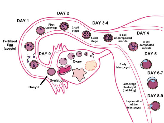
Figure 3.4. The germinal stage of human development begins with fertilization in a Fallopian tube and ends with implantation in the uterus.
The fertilized egg contains 46 chromosomes, 23 from each parent. In the few days after fertilization, cells divide, first gradually and then faster, through a process called mitosis, making an exact copy of all 46 chromosomes, dividing into two cells, and then repeating the process again, until all cells except those required for sexual reproduction are created. Sperm and ova, called gametes, are the cells for sexual reproduction. They are created through meiosis. The gametes duplicate, but then divide twice instead of once, making four cells with only 23 chromosomes, exactly half. When fertilization occurs, the sperm and ova combine to complete the 46 chromosomes, resulting in exactly half of the genetic material contributed from each biological parent.
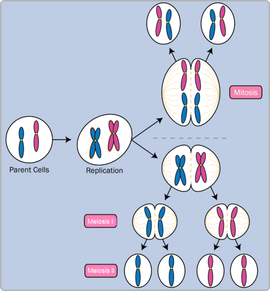
Figure 3.5. Diagram of cell division showing mitosis and meiosis.
3.4.1.2 Embryonic Period
The implantation of the blastocyst begins the next period, called the embryonic period. The implanted blastocyst, now a multicellular organism, is now referred to as an embryo (Figure 3.6). As cells divide they are beginning to differentiate during this period. Development occurs from head to toe, a pattern that will continue on into toddler years. Implantation is also what initiates the secretion of human chorionic gonadotrophic hormone (hCG). Secretion of hCG beginning in this 3rd or 4th week is the hormone detected by pregnancy tests. During this period, which is approximately 3 weeks post-fertilization, blood vessels are growing to form a structure called the placenta. The placenta is critical in the development of the fetus. This is where the exchange of nutrients, waste, hormones, and antibodies occurs between the mother and the developing fetus.
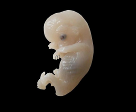
Figure 3.6. A human embryo.
About 20 percent of organisms fail during the embryonic period, usually due to chromosomal abnormalities, often before the mother even knows that she is pregnant. It is during this stage that the major structures of the body are taking form, making the embryonic period the time when the organism is most vulnerable to the greatest amount of damage if exposed to harmful substances. Prospective mothers are not often aware of the risks they introduce to the developing embryo during this time. The embryo is approximately 1 inch in length and weighs about 4 grams at the end of 8 weeks. The embryo can move and respond to touch at this time.
3.4.1.3 Fetal Period
From the 9th week until birth (which is approximately 40 weeks for a full-term pregnancy), the organism is referred to as a fetus. During this stage, the major structures are continuing to develop. By the 3rd month, the fetus has differentiated body parts including external genitalia. The fetus is about 3 in. long and weighs about 28 gm. Typical development in the following weeks including the development of hair, nails, and teeth, and the excretory and digestive systems will continue to develop.
Typical development during months 4–6 include the eyes becoming more sensitive to light and hearing development. The respiratory system continues to develop, and reflexes such as sucking, swallowing, and hiccupping, develop. The fetus also begins establishing sleep cycles at this time.
Between months 7–9, the fetus is primarily preparing for birth and viability outside of the womb. It is exercising its muscles and its lungs begin to expand and contract. The fetus gains about 5 lb and 7 in. during this last trimester of pregnancy, and during the 8th month, a layer of fat develops under the skin. This layer of fat serves as insulation and helps the baby regulate body temperature after birth.
At around 36 weeks the fetus is almost ready for birth. It weighs about 6 lb and is about 18.5 in. long. Typically by week 37 all of the fetus’ organ systems are developed enough that it could survive outside the mother’s uterus without many of the risks associated with premature birth or the need for resuscitation. The fetus continues to gain weight and grow in length until approximately 40 weeks. By then, the fetus has very little room to move around and birth becomes imminent. The progression through the stages is shown in Figure 3.7.

Figure 3.7. Critical Periods of Prenatal Development.
3.4.1.4 Prenatal Brain Development
Fetal brain development begins during the 3rd week of gestation with the differentiation of stem cells, which are capable of producing all the different cells that make up the brain. In the 9th week, the brain appears smooth but has further differentiated into the forebrain, midbrain, and hindbrain. Throughout the pregnancy, the smoothness will change and the brain will form many folds for the different regions of the brain (Konkel, 2018).
While the foundational development of the brain is assembled in gestation, brain function will change and develop after birth and for decades to come. Brain development during the fetal period involves neuron production, migration, and differentiation. From the early fetal period until midgestation, most of the 85 billion neurons have been generated and many have already migrated to their brain positions. Neurogenesis, or the formation of neurons, is largely completed after 5 months of gestation. One exception is in the hippocampus, which continues to develop neurons throughout life (Tobin, 2019). Neurons that form the neocortex begin lining up and communicating. This is the part of the brain related to sight, sound, language, spatial reasoning, and conscious thought (Stiles, 2010). Regions of the brain that contain the cell bodies are referred to as gray matter because they look gray in appearance. The axons that form the neural pathways make up the white matter because they are covered in myelin, a fatty substance that is white in appearance. Myelin aids in both the insulation and efficiency of neural transmission.
3.4.2 Licenses and Attributions for Conception and Prenatal Development
Embryonic and Fetal Development is shared under a CC BY-NC-SA license and was authored, remixed, and/or curated by Martha Lally & Suzanne Valentine-French.
“Prenatal Development” by Paris, Ricardo, Raymond, & Johnson is licensed under CC BY 3.0.
Embryology, Placenta by Herrick and Bordoni is licensed under CC BY 4.0.
Prenatal Development by Diana Lang; Martha Lally; Suzanne Valentine-French; Alisa Beyer; Julie Lazzara; and Naomi H. Dan Karami is licensed under a CC BY-NC-SA 4.0.
Figure 3.4. “Human Fertilization” by Ttrue12, Wikimedia Commons, is licensed under CC BY-SA 3.0.
Figure 3.5. “Mitosis vs. Meiosis” by Community College Consortium for Bioscience Credentials, Wikimedia Commons, is licensed under CC BY 3.0.
Figure 3.6. “Human Embryo” by Anatomist90, Wikimedia Commons, is licensed under CC BY-SA 3.0.
Figure 3.7. Image by Psyc 200 Lifespan Psychology. Authored by: Laura Overstreet. License under CC BY 4.0.