"

Lesson 3: A & P Terminology

Birds balancing on a branch

A&P Terminology

By the end of this lesson, students will be able to:

  • Locate various organs in the body using the basic anatomical terms for:
    • body position (anterior/posterior, supine/prone)
    • direction (superior/inferior, lateral/medial, proximal/distal,   superficial/deep)
    • plane (sagittal/frontal/transverse)
    • compartments (dorsal/ventral)
    • body regions
  • Identify the major body cavities, their membranes, and membrane layers

Introduction

Understanding anatomical terminology is essential for accurately describing the locations and relationships of structures in the human body. This module introduces key terms related to body position, directional references, planes, compartments, body cavities, membranes, and regional terminology.

Body Position Terms

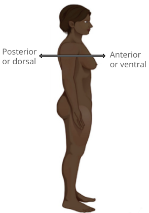
  • Anterior (ventral): Toward the front of the body
  • Posterior (dorsal): Toward the back of the body
  • Supine: Lying on the back, face upward
  • Prone: Lying on the stomach, face downward
Image of person looking to the right with arrows pointing anteriorly and posteriorly
Figure 1. Body Position. Anterior and Posterior

Directional Terms

  • Superior: Above or toward the head
  • Inferior: Below or toward the feet
  • Medial: Toward the midline of the body
  • Lateral: Away from the midline
  • Proximal: Closer to the point of attachment to the body
  • Distal: Further from the point of attachment to the body
  • Superficial: Closer to the surface
  • Deep: Further from the surface
This illustration shows two diagrams: one of a side view of a female and the other of an anterior view of a female. Each diagram shows directional terms using double-sided arrows. The cranial-distal arrow runs vertically behind the torso and lower abdomen. The cranial arrow is pointing toward the head while the caudal arrow is pointing toward the tail bone. The posterior/anterior arrow is running horizontally through the back and chest. The posterior or dorsal arrow is pointing toward the back while the anterior, or ventral arrow, is pointing toward the abdomen. On the anterior view, the proximal/distal arrow is on the right arm. The proximal arrow is pointing up toward the shoulder while the distal arrow is pointing down toward the hand. The lateral-medial arrow is a horizontal arrow on the abdomen. The medial arrow is pointing toward the navel while the lateral arrow is pointing away from the body to the right. Right refers to the right side of the woman’s body from her perspective while left refers to the left side of the woman’s body from her perspective.
Figure 2. Directional Terms Applied to the Human Body. Paired directional terms are shown as applied to the human body.

Body Planes

  • Sagittal plane: Divides the body into left and right sections
  • Frontal (coronal) plane: Divides the body into front and back sections
  • Transverse plane: Divides the body into top and bottom sections
  • Oblique plane: A diagonal section through the body
This illustration shows a female viewed from her right, front side. The anatomical planes are depicted as blue rectangles passing through the woman’s body. The frontal or coronal plane enters through the right side of the body, passes through the body, and exits from the left side. It divides the body into front (anterior) and back (posterior) halves. The sagittal plane enters through the back and emerges through the front of the body. It divides the body into right and left halves. The transverse plane passes through the body perpendicular to the frontal and sagittal planes. This plane is a cross section which divides the body into upper and lower halves.
Figure 3. Planes of the Body. The three planes most commonly used in anatomical and medical imaging are the sagittal, frontal (or coronal), and transverse planes.

Body Cavities and Membranes

  • Dorsal body cavity: Contains the cranial and spinal cavities
  • Ventral body cavity: Contains the thoracic and abdominopelvic cavities
This illustration shows a lateral and anterior view of the body and highlights the body cavities with different colors. The cranial cavity is a large, bean-shaped cavity filling most of the upper skull where the brain is located. The vertebral cavity is a very narrow, thread-like cavity running from the cranial cavity down the entire length of the spinal cord. Together the cranial cavity and vertebral cavity can be referred to as the dorsal body cavity. The thoracic cavity consists of three cavities that fill the interior area of the chest. The two pleural cavities are situated on both sides of the body, anterior to the spine and lateral to the breastbone. The superior mediastinum is a wedge-shaped cavity located between the superior regions of the two thoracic cavities. The pericardial cavity within the mediastinum is located at the center of the chest below the superior mediastinum. The pericardial cavity roughly outlines the shape of the heart. The diaphragm divides the thoracic and the abdominal cavities. The abdominal cavity occupies the entire lower half of the trunk, anterior to the spine. Just under the abdominal cavity, anterior to the buttocks, is the pelvic cavity. The pelvic cavity is funnel shaped and is located inferior and anterior to the abdominal cavity. Together the abdominal and pelvic cavity can be referred to as the abdominopelvic cavity while the thoracic, abdominal, and pelvic cavities together can be referred to as the ventral body cavity.
Figure 4. Dorsal and Ventral Body Cavities The ventral cavity includes the thoracic and abdominopelvic cavities and their subdivisions. The dorsal cavity includes the cranial and spinal cavities.
  • Membranes: A tissue membrane is a thin layer or sheet of cells that covers the outside of the body, the organs, internal passageways that lead to the exterior of the body, and the lining of the moveable joint cavities. There are two basic types of tissue membranes: connective tissue and epithelial membranes.  Mucous membranes, serous membranes, and cutaneous membranes are all epithelial membranes because they are derived from epithelium.  The synovial membrane is a connective tissue membrane.  Here is a list of their functions and locations:
    • Mucous membranes: Line cavities open to the exterior (e.g., digestive and respiratory tracts)
    • Serous membranes: Line body cavities closed to the exterior and consist of two layers:
      • Visceral layer: Covers internal organs
      • Parietal layer: Lines the cavity walls
    • Cutaneous membrane: The skin
    • Synovial membrane: Lines joint cavities and produces synovial fluid
This illustrations shows the silhouette of a human female from an anterior view. Several organs are showing in her neck, thorax, abdomen left arm and right leg. Text boxes point out and describe the mucous membranes in several different organs. The topmost box points to the mouth and trachea. It states that mucous membranes line the digestive, respiratory, urinary and reproductive tracts. They are coated with the secretions of mucous glands. The second box points to the outside edge of the lungs as well as the large intestine and states that serous membranes line body cavities that are closed to the exterior of the body, including the peritoneal, pleural and pericardial cavities. The third box points to the skin of the hand. It states that cutaneous membrane, also known as the skin, covers the body surface. The fourth box points to the right knee. It states that synovial membranes line joint cavities and produce the fluid within the joint.
Figure 5. Tissue Membranes The two broad categories of tissue membranes in the body are (1) connective tissue membranes, which include synovial membranes, and (2) epithelial membranes, which include mucous membranes, serous membranes, and the cutaneous membrane, in other words, the skin.

Body Regions

  • Head and Neck: Cephalic, cervical, cranial, facial, occipital, orbital
  • Torso: Thoracic, pectoral, abdominal, lumbar, umbilical
  • Upper Limb: Acromial, brachial, antecubital, carpal, digital
  • Lower Limb: Coxal, femoral, patellar, crural, tarsal, plantar
This illustration shows an anterior and posterior view of the human body. The cranial region encompasses the upper part of the head while the facial region encompasses the lower half of the head beginning below the ears. The eyes are referred to as the ocular region. The cheeks are referred to as the buccal region. The ears are referred to as the auricle or otic region. The nose is referred to as the nasal region. The chin is referred to as the mental region. The neck is referred to as the cervical region. The trunk of the body contains, from superior to inferior, the thoracic region encompassing the chest, the mammary region encompassing each breast, the abdominal region encompassing the stomach area, the coxal region encompassing the belt line, and the pubic region encompassing the area above the genitals. The umbilicus, or naval, is located at the center of the abdomen. The pelvis and legs contain, from superior to inferior, the inguinal or groin region between the legs and the genitals, the pubic region surrounding the genitals, the femoral region encompassing the thighs, the patellar region encompassing the knee, the crural region encompassing the lower leg, the tarsal region encompassing the ankle, the pedal region encompassing the foot and the digital/phalangeal region encompassing the toes. The great toe is referred to as the hallux. The regions of the upper limbs, from superior to inferior, are the axillary region encompassing the armpit, the brachial region encompassing the upper arm, the antecubital region encompassing the front of the elbow, the antebrachial region encompassing the forearm, the carpal region encompassing the wrist, the palmar region encompassing the palm, and the digital/phalangeal region encompassing the fingers. The thumb is referred to as the pollux. The posterior view contains, from superior to inferior, the cervical region encompassing the neck, the dorsal region encompassing the upper back and the lumbar region encompassing the lower back. The regions of the back of the arms, from superior to inferior, include the cervical region encompassing the neck, acromial region encompassing the shoulder, the brachial region encompassing the upper arm, the olecranal region encompassing the back of the elbow, the antebrachial region encompasses the back of the arm, and the manual region encompassing the palm of the hand. The posterior regions of the legs, from superior to inferior, include the gluteal region encompassing the buttocks, the femoral region encompassing the thigh, the popliteus region encompassing the back of the knee, the sural region encompassing the back of the lower leg, and the plantar region encompassing the sole of the foot. Some regions are combined into larger regions. These include the trunk, which is a combination of the thoracic, mammary, abdominal, naval, and coxal regions. The cephalic region is a combination of all of the head regions. The upper limb region is a combination of all of the arm regions. The lower limb region is a combination of all of the leg regions.
Figure 6. Regions of the Human Body The human body is shown in anatomical position in an (a) anterior view and a (b) posterior view. The regions of the body are labeled in boldface.

Allied Health Anatomy: Examination Methods

  • Inspection: Visual observation
  • Dissection: Cutting and separation of tissues to reveal relationships
  • Palpation: Feeling structures with fingertips
  • Auscultation: Listening to body sounds using a stethoscope
  • Percussion: Tapping with fingers or a small instrument to detect differences in density
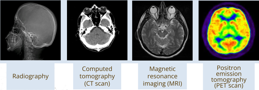
  • Medical Imaging:
    • X-ray
    • CT scan
    • MRI
    • PET scan
This figure shows four images from different techniques: X-ray, CT scan, MRI, and PET scan
Figure 6. Medical Imaging. This figure shows images from X-ray, CT scan, Magnetic Resonance Imaging (MRI), and Positron Emission Tomography (PET) scan

Anatomical Variation

  • Individual differences exist in body structures, such as extra muscles, missing organs, or accessory spleens.
  • Physiology, or function, is often more variable than anatomy.
Diagram of a horseshoe kidney on the left and typical kidneys on the right.
Figure 7. Anatomical Variation. Diagram of a horseshoe kidney on the left and typical kidneys on the right.

Summary

Anatomical terminology provides a universal language for describing body structures, essential for medical professionals and students. Understanding these terms ensures precise communication and enhances the study of anatomy and physiology.


Watch this lesson video walking you through the Module 1 Lesson 3 A & P Terminology (PDF) slides.


Practice Questions

Use these practice questions to assess your knowledge before you move on to the next section.

License

Icon for the Creative Commons Attribution-ShareAlike 4.0 International License

Introduction to Human Anatomy & Physiology: A Multilingual Approach Copyright © by Rachel Thwing; Hugh Jarrard; Ann DeChenne; Kiana Pigao; and Zach Ellsworth is licensed under a Creative Commons Attribution-ShareAlike 4.0 International License, except where otherwise noted.

Share This Book