"

Lesson 1: Functions of the Integumentary System

Skin model
Model of Human Skin By Scivit, licensed under CC BY-SA 4.0. Fat cells from the hypodermis are in blue. Blood vessels are red, nerve tissue is green, while hairs and glands are yellow. The connective tissue of the hypodermis and dermis is brown. The epidermis on top is not shown.

Functions of the Integumentary System

By the end of this lesson, students will be able to:

  • Explain the functions of the integumentary system.
  • Identify the principal structures and layers of human skin.
  • Identify Accessory Structures and their function.

Introduction

The integumentary system is a vital and complex organ system that serves as the body’s primary interface with the external environment. In this lesson, we will explore the structure and function of the integumentary system. Our primary objectives are to explain its key functions, identify the principal structures and layers of human skin, and describe the structure and function of accessory structures found in and on human skin.

Functions of the Integumentary System

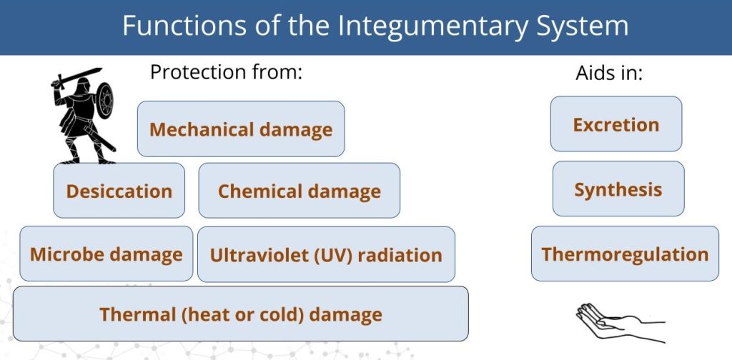
The integumentary system acts first and foremost as a protective shield, guarding the body from mechanical, chemical, microbial, thermal, and ultraviolet (UV) damage. It also prevents desiccation, plays roles in excretion and vitamin D synthesis, and helps regulate body temperature. See Figure 1.

Image shows the protective functions of skin as if they are bricks with a viking on top to symbolize protection. Skin protects from various types of damage. The functions of skin in excretion, synthesis, and thermoregulation are symbolized with a helping hand.
Figure 1. Functions of Integument. The skin protects from various types of damage and aids in excretion, synthesis, and thermoregulation.

Protection from External Damage

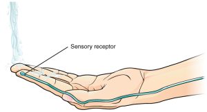
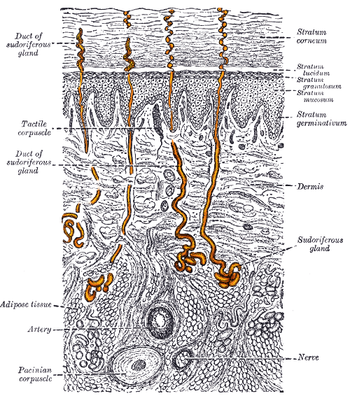
Mechanical Protection: The skin is composed of tough, tightly bound cells rich in keratin—a protein that reinforces the skin’s structure. Beneath the skin lies a layer of adipose tissue that cushions mechanical blows. Sensory receptors embedded in the skin detect pressure and pain, alerting the nervous system to potential injury. See Figure 2.

Confocal microscopy of keratin filaments. Fibers appear like red strings being pulled apart
Figure 2. Keratin fibers form a water-resistant physical barrier on the skin surface. Keratin F9, CC BY-SA 3.0, via Wikimedia Commons

Chemical Protection: The outermost keratinized layer of skin is relatively impermeable to many aqueous solutions, including acids and bases. The outermost layer of cells are packed tightly, and contain a specialized arrangement of lipids and cells that prevent excessive water loss and entry. See Figure 3. Pain receptors help detect chemical threats, prompting quick behavioral responses.

water drops beaded on skin
Figure 3. Water Drops beaded on the skin demonstrate the water resistance of the skin. By Иван Стойков, CC BY-SA 3.0

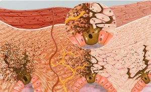
Microbial Protection: The skin serves as a physical barrier against pathogens. Its secretions create an acidic environment (acid mantle) that inhibits microbial growth. If pathogens do breach the surface, immune cells located just beneath the skin can phagocytize and destroy them. See Figure 4.

Phagocytosis is the process in which a cell engulfs a particle, digests it, and expels the waste products.Process of phagocytosis: 1. A particle is ingested by a phagocyte after antigens are recognized which results in the formation of a phagosome. 2. The fusion of lysosomes with the phagosome creates a phagolysosome. The particle is broken down by the digestive enzymes found in the lysosomes.
Figure 4. Phagocytosis is the process in which a cell engulfs a particle, digests it, and expels the waste products. Process of phagocytosis: 1. A particle is ingested by a phagocyte which results in the formation of a phagosome inside the cell. 2. Fusion with a lysosome causes the particle to be broken down by the digestive enzymes found in the lysosomes. 3. The resulting waste material is discharged by exocytosis. Image By Mango Slices, CC BY-SA 4.0, via Wikimedia Commons.

UV Radiation Protection: Specialized cells called melanocytes, located in deeper layers of the skin, produce melanin. Melanin is a dark pigment that absorbs UV rays, shielding underlying cells from DNA damage. See Figure 5.

3D Cross-section showing skin tone becoming darker due to the production of more melanin to overcome the damage of melanin caused by sun tanning
Figure 5. Melanin and Melanocytes. This is a 3D Cross-section showing skin tone becoming darker due to the production of more melanin to overcome the damage of melanin caused by sun tanning. Image by Scientific Animations, CC BY-SA 4.0

Thermal Protection: Heat, cold, and pain receptors help detect environmental temperature extremes, prompting protective behaviors. The skin also contributes to thermoregulation by controlling blood flow through capillary networks near its surface. See Figure 6.

Hand feeling cold water. Protection from Thermal Damage.Skin detects hot and cold stimuli to help the body avoid thermal injury.
Figure 6. Protection from Thermal Damage. Skin detects hot and cold stimuli to help the body avoid thermal injury. From Anatomy and Physiology by OpenStax, licensed under CC BY 4.0

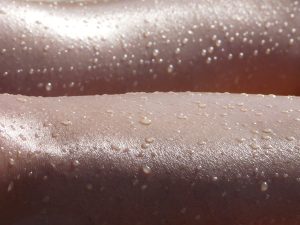
Prevention of Desiccation: The keratinized outer layer contains glycolipids that repel water, preventing dehydration by keeping internal moisture in and external moisture out.

Additional Functions

Excretion

The skin helps eliminate nitrogenous wastes such as urea and uric acid through the sweat glands. Though the kidneys are the primary organ of excretion, the integumentary system contributes by secreting waste in sweat. See Figure 7.

sectional view of skin with sweat glands highlighted
Figure 7. Excretion of Nitrogenous Waste. Sweat glands excrete small amounts of metabolic waste products. Sectional View of Skin with Sweat Glands Highlighted By Henry Gray – Gray’s Anatomy, Public Domain.

Synthesis of Vitamin D

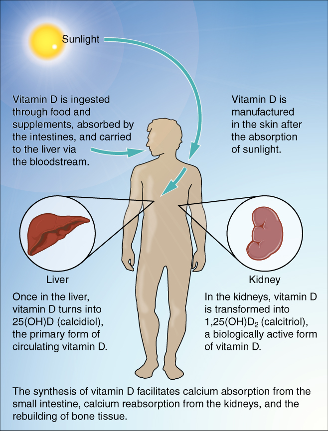
When exposed to UV rays, cholesterol molecules in the skin are converted into vitamin D. This vitamin is essential for calcium absorption and overall metabolic function, making the skin an important site of chemical synthesis.

UV exposure enables the skin to convert cholesterol to vitamin D.
Figure 8. Synthesis of Vitamin D. By OpenStax College – Anatomy and Physiology, Connexions Web site. CC BY 3.0, via Wikimedia Commons

Thermoregulation

The integumentary system helps maintain stable internal temperature. When the body overheats, blood vessels in the dermis dilate, allowing warm blood to reach the skin surface where heat can dissipate. This causes the skin to appear flushed or red. Conversely, in cold conditions, blood vessels constrict, reducing blood flow near the surface and preserving body heat—resulting in paler or bluish skin.

Part A is a photo of a man skiing with several snow-covered trees in the background. Part B is a diagram with a right and left half. The left half is titled “ Heat is retained by the body,” while the right half is titled “Heat loss through radiation and convection.” Both show blood flowing from an artery through three capillary beds within the skin. The beds are arranged vertically, with the topmost bed located along the boundary of the dermis and epidermis. The bottommost bed is located deep in the hypodermis. The middle bed is evenly spaced between the topmost and bottommost beds. In each bed, oxygenated blood (red) enters the bed on the left and deoxygenated blood (blue) leaves the bed on the right. The left diagram shows a picture of snowflakes above the capillary beds, indicating that the weather is cold. Blood is only flowing through the deepest of the three capillary beds, as the upper beds are closed off to reduce heat loss from the outer layers of the skin. The right diagram shows a picture of the sun above the capillary beds, indicating that the weather is hot. Blood is flowing through all three capillary beds, allowing heat to radiate out of the blood, increasing heat loss. Part C is a photo of a man running through a forested trail on a summer day.
Figure 9. During strenuous physical activities, such as skiing (a) or running (c), the dermal blood vessels dilate and sweat secretion increases (b). These mechanisms prevent the body from overheating. In contrast, the dermal blood vessels constrict to minimize heat loss in response to low temperatures (b). By OpenStax College – Anatomy and Physiology, 2e

Structure of the Skin

The skin has two primary layers: the epidermis and the dermis, with a third supportive layer beneath called the hypodermis. The epidermis is made of closely packed epithelial cells. The dermis is made of dense, irregular connective tissue that houses blood vessels, hair follicles, sweat glands, and other structures. Beneath the dermis lies the hypodermis, which is composed mainly of loose connective and fatty tissues. See Figure 10.


This illustration shows a cross section of skin tissue. The outermost layer is called the epidermis, and occupies one fifth of the cross section. Several hairs are emerging from the surface. The epidermis dives around one of the hairs, forming a follicle. The middle layer is called the dermis, which occupies four fifths of the cross section. The dermis contains an erector pilli muscle connected to one of the follicles. The dermis also contains an eccrine sweat gland, composed of a bunch of tubules. One tubule travels up from the bunch, through the epidermis, opening onto the surface a pore. There are two string-like nerves travelling vertically through the dermis. The right nerve is attached to a Pacinian corpuscle, which is a yellow structure consisting of concentric ovals similar to an onion. The lowest level of the skin, the hypodermis, contains fatty tissue, arteries, and veins. Blood vessels travel from the hypodermis and connect to hair follicles and erector pilli muscle in the dermis.
Figure 10. Layers of the Skin. The skin includes the epidermis, dermis, and subcutaneous hypodermis. By OpenStax College – Anatomy and Physiology, 2e


Epidermis

The epidermis is the outermost layer, composed of stratified squamous epithelium. It contains keratinocytes, which produce keratin and form a waterproof barrier. These cells originate in the deepest layer and move upward as they mature, eventually dying and being sloughed off.

This illustration shows a cross section of the epidermis. The cells of the innermost layer, the stratum basale, are large and have a purple nucleus. The stratum basale curls around the dermis, which projects into the epidermis. The stratum basale contains four layers of large, triangle-shaped keratinocytes. Fibers are visible within the spaces between keratinocytes in the stratum basale. A melanocyte is also present in this layer. The melanocyte possesses finger-like projections extending from its main cell body. The projections branch through the extracellular spaces between nearby keratinocytes. Above the stratum basale is the stratum spinosum which consists of 8 layers of oval-shaped keratinocytes. The nucleus is present in these keratinocytes, but has faded to a lighter purple. The stratum granulosum contains five layers of keratinocytes, each containing spots in its cytoplasm, labeled the lamellar granules. The stratum lucidium contains 4 layers of diamond-shaped cells with no nucleus. The stratum corneum contains 9 layers of keratinocytes with no nucleus , nor cytoplasm. A few of the cells in the topmost layer of the stratum corneum are flaking off from the skin.
Figure 11. Layers of the Epidermis. Five strata protect the body and support cell renewal in thick skin. By OpenStax College – Anatomy and Physiology, 2e

The layers of the epidermis, from deepest to most superficial, are:

  • Stratum basale: Contains basal cells that continually divide to produce new keratinocytes.

  • Stratum spinosum: Cells are interconnected by desmosomes, which give the layer a spiny appearance.

  • Stratum granulosum: Cells accumulate keratin granules, preparing for their eventual death.

  • Stratum lucidum (found only in thick skin): A thin, translucent layer present in the palms and soles.

  • Stratum corneum: The outermost layer composed of dead, flattened, keratin-filled cells that are regularly shed.

Other important epidermal cells include:

  • Merkel cells: Sensory cells that detect touch.

  • Langerhans cells: Immune cells that ingest foreign microbes.

  • Melanocytes: Pigment-producing cells that help block UV radiation.

Dermis

Beneath the epidermis lies the dermis, made of connective tissue rich in collagen and elastin fibers. It contains blood vessels, nerve endings, glands, and hair follicles. See Figure 12.

The dermis has two layers with a rich sensory and vascular network:

  • Papillary layer: The superficial region composed of dermal papillae—finger-like projections that increase surface area and strengthen attachment to the epidermis.

  • Reticular layer: The deeper region composed of dense irregular connective tissue that provides strength and flexibility. It houses most of the skin’s accessory structures.

This micrograph shows layers of skin in a cross section. The papillary layer of the dermis extends between the downward fingers of the darkly stained epidermis. The papillary layer appears finer than the reticular layer, consisting of smaller, densely-packed fibers. The reticular layer is three times thicker than the papillary layer and contains larger, thicker fibers. The fibers seem more loosely packed than those of the papillary layer, with some separated by empty spaces. Both layers of the dermis contain cells with darkly stained nuclei.
Figure 12. Layers of the Dermis. This stained slide shows the two components of the dermis—the papillary layer and the reticular layer. Both are made of connective tissue with fibers of collagen extending from one to the other, making the border between the two somewhat indistinct.  LM × 10. Credit: modification of work by “kilbad”/Wikimedia Commons By OpenStax College – Anatomy and Physiology, 2e

Hypodermis

The hypodermis, or subcutaneous layer, is not part of the skin.  It lies beneath the dermis and contains adipose tissue and connective tissue, providing insulation, energy storage, and cushioning. Below the hypodermis, bone and skeletal muscle are typically found.

Accessory Structures of the Skin

The integumentary system also includes several important accessory structures: hair, nails, and glands.

Hair

Hair is composed of keratinized cells and serves several functions, including thermoregulation, sensory detection, and protection. The visible part that extends above the skin is the shaft, while the root is the base anchored in the skin, and the matrix is the region of active cell division that produces the hair. The arrector pili muscle is the smooth muscle attached to the hair follicle that contracts to raise the hair shaft, causing goosebumps. See Figure 13.

This diagram shows a cross section of the skin containing a hair follicle. The follicle is teardrop shaped. Its enlarged base, labeled the hair bulb, is embedded in the hypodermis. The outermost layer of the follicle is the epidermis, which invaginates from the skin surface to envelope the follicle. Within the epidermis is the outer root sheath, which is only present on the hair bulb. It does not extend up the shaft of the hair. Within the outer root sheath is the inner root sheath. The inner root sheath extends about half of the way up the hair shaft, ending midway through the dermis. The hair matrix is the innermost layer. The hair matrix surrounds the bottom of the hair shaft where it is embedded within the hair bulb. The hair shaft, in itself, contains three layers: the outermost cuticle, a middle layer called the cortex, and an innermost layer called the medulla.
Figure 13. Hair follicles originate in the epidermis and have many different parts. They provide insulation, sensory input, and protection. By OpenStax College – Anatomy and Physiology, 2e

The functions of hair include:

  • Sensory reception

  • Protection from UV radiation

  • Thermoregulation (via arrector pili muscle contraction)

Nails

Nails are also made of keratinized cells and provide protection to the fingertips and aid in grasping small objects. The nail body lies on top of the nail bed, and the cuticle protects the growth area at the base of the nail. See Figure 14.

These two images show anatomy of the fingernail region. The top image shows a dorsal view of a finger. The proximal nail fold is the part underneath where the skin of the finger connects with the edge of the nail. The eponychium is a thin, pink layer between the white proximal edge of the nail (the lunula), and the edge of the finger skin. The lunula appears as a crescent-shaped white area at the proximal edge of the pink-shaded nail. The lateral nail folds are where the sides of the nail contact the finger skin. The distal edge of the nail is white and is called the free edge. An arrow indicates that the nail grows distally out from the proximal nail fold. The lower image shows a lateral view of the nail bed anatomy. In this view, one can see how the edge of the nail is located just proximal to the nail fold. This end of the nail, from which the nail grows, is called the nail root.
Figure 14. Nails. The nail is an accessory structure of the integumentary system. Keratinized nail plates provide protection and aid in object manipulation. By OpenStax College – Anatomy and Physiology, 2e

Glands

There are two primary types of sweat glands and one type of oil gland:

  • Sebaceous glands: Produce sebum, a fatty substance that lubricates and waterproofs the skin and hair.

  • Eccrine sweat glands: Widely distributed; produce watery, odorless sweat that evaporates to cool the body.

  • Apocrine sweat glands: Found in the armpits and genital regions, they secrete a thicker, milky sweat that contributes to body odor and may have once played a role in pheromone-based communication.

Both eccrine and apocrine glands contribute to the excretion of metabolic waste such as urea. See Figure 15.

This illustration shows a cross section of skin tissue. The outermost layer is called the epidermis, and occupies one fifth of the cross section. Several hairs are emerging from the surface. The epidermis dives around one of the hairs, forming a follicle. The middle layer is called the dermis, which occupies four fifths of the cross section. The dermis contains an erector pilli muscle connected to one of the follicles. The dermis also contains an eccrine sweat gland, composed of a bunch of tubules. One tubule travels up from the bunch, through the epidermis, opening onto the surface a pore. There are two string-like nerves travelling vertically through the dermis. The right nerve is attached to a Pacinian corpuscle, which is a yellow structure consisting of concentric ovals similar to an onion. The lowest level of the skin, the hypodermis, contains fatty tissue, arteries, and veins. Blood vessels travel from the hypodermis and connect to hair follicles and erector pilli muscle in the dermis.
Figure 15. Skin Glands. Eccrine and apocrine sweat glands serve roles in thermoregulation, excretion, and social signaling. By OpenStax College – Anatomy and Physiology, 2e

Summary

In this lesson, we explored the essential components and functions of the integumentary system. We discussed its role in protection, excretion, thermoregulation, and vitamin synthesis. We examined the structural layers of the skin, including the epidermis, dermis, and hypodermis, and learned about accessory structures such as hair, nails, and glands. This system is both the body’s first line of defense and a key regulator of internal balance and homeostasis.


Watch this lesson video, walking you through the Module 2 Lesson 1 Integumentary Structure & Function slides.


Practice Questions

Use these practice questions to assess your knowledge before you move on to the next section.


License

Icon for the Creative Commons Attribution-ShareAlike 4.0 International License

Introduction to Human Anatomy & Physiology: A Multilingual Approach Copyright © by Rachel Thwing; Hugh Jarrard; Ann DeChenne; Kiana Pigao; and Zach Ellsworth is licensed under a Creative Commons Attribution-ShareAlike 4.0 International License, except where otherwise noted.

Share This Book