"

Lesson 1: Levels of Organization and Organ Systems Overview

Birds balancing on a branch

Levels of Structural Organization and Organ Systems

By the end of this lesson, students will be able to:

  • Define, in order from simplest (chemical) to most complex (organismal), the major levels of structural organization in the human organism.
  • Utilize systemic and regional approaches to relate gross anatomical structures to their functions.
  • Explain how the cells and forms of the tissues contribute to their function in the body.
  • Identify the different tissue types (nervous, muscle, connective, epithelial, and membrane) by microscopic methods.

Introduction

The human body is organized into hierarchical levels of structural complexity, from the simplest chemical structures to the entire organism. This organization supports the proper function and regulation of physiological processes, ensuring homeostasis and survival.

Levels of Structural Organization

The following hierarchical levels of structural complexity, are arranged below in Figure 1 from the simplest (top) to the most complex (base).  As we gain complexity, properties emerge, demonstrating that the sum is greater than its parts.

  1. Atoms – The basic units of matter that combine to form molecules.
  2. Cells – The smallest functional units of life, composed of molecules.
  3. Tissues – Groups of similar cells working together to perform a common function.
  4. Organs – Structures of multiple tissue types working together for a specific function.
  5. Organ Systems – Groups of organs that work collaboratively to perform bodily functions.
  6. Organism – The complete human body functioning as a whole.
This illustration shows biological organization as a pyramid. The chemical level is at the apex of the pyramid where atoms bond to form molecules with three dimensional structures. An example is shown with two white hydrogen atoms bonding to a red oxygen atom to create water. The next level down on the pyramid is the cellular level, as illustrated with a long, tapered, smooth muscle cell. At this level, a variety of molecules combine to form the interior fluid and organelles of a body cell. The next level down is the tissue level. A community of similar cells forms body tissue. The example given here is a section of smooth muscle tissue, which contains many smooth muscle cells closely bound side by side. The next level down is the organ level, as illustrated with the bladder and urethra. The bladder contains smooth muscle while the urethra contains skeletal muscle. These are both examples of muscle tissues. The next level down is the organ system level, as illustrated by the entire urinary system containing the kidney, ureters, bladder and urethra. At this level, two or more organs work closely together to perform the functions of a body system. At the base of the pyramid is the organismal level illustrated with a woman drinking water. At this level, many organ systems work harmoniously together to perform the functions of an independent organism.
Figure 1. Levels of Structural Organization of the Human Body. The body’s organization is often discussed in six levels of increasing complexity, from the smallest chemical building blocks to a unique human organism.

Anatomy and Organization of the Human Body

Gross Anatomy versus Microscopic Anatomy

  • Gross Anatomy refers to structures visible to the naked eye, studied via systemic, regional, and surface approaches. See Figure 2.
  • Histology examines tissues and cells under a microscope to understand their organization and function. See Figure 3.
This diagram shows the digestive system of a human being, with the major organs labeled.
Figure 2. Components of the Digestive System All digestive organs play integral roles in the life-sustaining process of digestion.
This diagram shows the histological cross-section of the stomach. The left panel shows the stomach and the center panel shows a magnified view of a small region including the epithelium and the gastric glands. The right panel shows a further magnification of the mucosa and the different cell types are labeled.
Figure 3. Histology of the Stomach The stomach wall is adapted for the functions of the stomach. In the epithelium, gastric pits lead to gastric glands that secrete gastric juice. The gastric glands (one gland is shown enlarged on the right) contain different types of cells that secrete various enzymes, including hydrochloride acid, which activates the protein-digesting enzyme pepsin.

Tissue Types

Epithelial tissue, also called epithelium, refers to the sheets of cells that cover exterior surfaces of the body, line internal cavities and passageways, and form certain glands. Connective tissue, as its name implies, binds the cells and organs of the body together and functions in the protection, support, and integration of all parts of the body. Muscle tissue is excitable, responding to stimulation and contracting to provide movement, and occurs in three major types: skeletal (voluntary) muscle, smooth muscle, and cardiac muscle in the heart. Nervous tissue is also excitable, allowing the propagation of electrochemical signals through nerve impulses that communicate between different body regions.

This diagram shows the silhouette of a female surrounded by four micrographs of tissue. Each micrograph has arrows pointing to the organs where that tissue is found. The upper left micrograph shows nervous tissue that is whitish with several large, purple, irregularly-shaped neurons embedded throughout. Nervous tissue is found in the brain, spinal cord and nerves. The upper right micrograph shows muscle tissue that is red with elongated cells and prominent, purple nuclei. Cardiac muscle is found in the heart. Smooth muscle is found in muscular internal organs, such as the stomach. Skeletal muscle is found in parts that are moved voluntarily, such as the arms. The lower left micrograph shows epithelial tissue. This tissue is purple with many round, purple cells with dark purple nuclei. Epithelial tissue is found in the lining of GI tract organs and other hollow organs such as the small intestine. Epithelial tissue also composes the outer layer of the skin, known as the epidermis. Finally, the lower right micrograph shows connective tissue, which is composed of very loosely packed purple cells and fibers. There are large open spaces between clumps of cells and fibers. Connective tissue is found in the leg within fat and other soft padding tissue as well as bones and tendons.
Figure 4. Four Types of Tissue: Body The four types of tissues are exemplified in nervous tissue, stratified squamous epithelial tissue, cardiac muscle tissue, and connective tissue. (Micrographs provided by the Regents of University of Michigan Medical School © 2012)

Epithelial Tissue

    • Locations: Covers surfaces, lines cavities.
    • Functions: Protection, absorption, secretion, excretion, filtration.
    • Characteristics: Tightly packed cells, rapid regeneration, presence of a free surface.
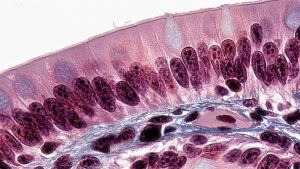
This photomicrograph shows the simple columnar epithelium lining the intestine.  We see column-shaped cells with microvilli brush borders to increase surface area for secretion and absorption.  The microvilli face the lumen of the tube so that they come into direct contact with foodstuffs.
Figure 5. Epithelial Tissue.  This photomicrograph shows the simple columnar epithelium lining the intestine.  We see column-shaped cells with microvilli brush borders to increase surface area for secretion and absorption.  The microvilli face the lumen of the tube so that they come into direct contact with foodstuffs.


Connective Tissue

    • Locations: Found throughout the body.
    • Functions: Binds structures, provides support, stores energy, and transports substances.
    • Characteristics: Composed of few cells within an extracellular matrix (EM), slow replication.
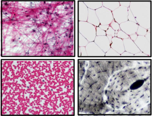
This photomicrograph shows loose areolar connective tissue in the upper left frame, adipose in the upper right frame,  blood in the lower left frame, and bone in the lower right frame. 
Figure 6. Connective Tissue.  This photomicrograph shows loose areolar connective tissue in the upper left frame, adipose in the upper right frame,  blood in the lower left frame, and bone in the lower right frame.

Muscular Tissue

    • Locations: Attached to bones, within the heart, in the walls of hollow organs.
    • Functions: Enables movement and stability.
    • Characteristics: Highly cellular, vascular, capable of contraction.
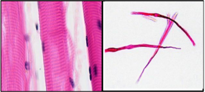
This photomicrograph shows skeletal muscle tissue in the left panel and smooth muscle tissue teased in the right panel. 
Figure 7. Muscle Tissue.  This photomicrograph shows skeletal muscle tissue in the left panel and smooth muscle tissue teased in the right panel.

Nervous Tissue

    • Locations: Brain, spinal cord, nerves.
    • Functions: Coordinates bodily functions through neurons and neuroglia.
    • Characteristics: Avascular, composed of neurons with axons and dendrites.
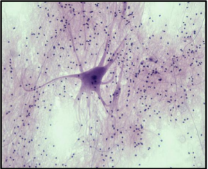
This photomicrograph shows nervous tissue.
Figure 8. Nervous Tissue.  This photomicrograph shows nervous tissue. The pyramid-shaped cell body of a neuron is stained darkly. Outgrowths from the cell body could be dendrites or axons. Glial cells are small, darkly stained cells surrounding the neuron.

Overview of Organ Systems

The next level of organization is the organ, where several types of tissues come together to form a working unit. Just as knowing the structure and function of cells helps you study tissues, knowledge of tissues will help you understand how organs function. The epithelial and connective tissues are discussed in detail in this chapter. Muscle and nervous tissues will be discussed only briefly in this chapter.

The human body consists of multiple organ systems, each performing specialized functions essential for survival and homeostasis.

This illustration shows eight silhouettes of a human female, each showing the components of a different organ system. The integumentary system encloses internal body structures and is the site of many sensory receptors. The integumentary system includes the hair, skin, and nails. The skeletal system supports the body and, along with the muscular system, enables movement. The skeletal system includes cartilage, such as that at the tip of the nose, as well as the bones and joints. The muscular system enables movement, along with the skeletal system, but also helps to maintain body temperature. The muscular system includes skeletal muscles, as well as tendons that connect skeletal muscles to bones. The nervous system detects and processes sensory information and activates bodily responses. The nervous system includes the brain, spinal cord, and peripheral nerves, such as those located in the limbs. The endocrine system secretes hormones and regulates bodily processes. The endocrine system includes the pituitary gland in the brain, the thyroid gland in the throat, the pancreas in the abdomen, the adrenal glands on top of the kidneys, and the testes in the scrotum of males as well as the ovaries in the pelvic region of females. The cardiovascular system delivers oxygen and nutrients to the tissues as well as equalizes temperature in the body. The cardiovascular system includes the heart and blood vessels.
Figure 9. Organ Systems of the Human Body. Organs that work together are grouped into organ systems.
  1. Integumentary System see Figure 9
    • Organs: Skin, hair, nails, glands.
    • Functions: Protection, water retention, thermoregulation, vitamin D synthesis.
  2. Skeletal System –see Figure 9
    • Organs: Bones, cartilage, ligaments.
    • Functions: Support, movement, blood formation, mineral storage.
  3. Muscular System –see Figure 9
    • Organs: Skeletal muscles.
    • Functions: Movement, stability, heat production.
  4. Nervous Systemsee Figure 9
    • Organs: Brain, spinal cord, nerves.
    • Functions: Rapid internal communication, motor control, sensory perception.
  5. Endocrine System-see Figure 9
    • Organs: Glands (pituitary, thyroid, adrenal, pancreas, etc.).
    • Functions: Hormone production and regulation of body functions.
  6. Cardiovascular System– see Figure 9
    • Organs: Heart, blood vessels.
    • Functions: Circulation of blood, nutrient and gas transport.
  7. Lymphatic System– see Figure 10
    • Organs: Lymph nodes, spleen, tonsils.
    • Functions: Immune response, fluid balance.
  8. Respiratory System– see Figure 10
    • Organs: Lungs, trachea, bronchi.
    • Functions: Oxygen intake, carbon dioxide expulsion.
  9. Digestive System– see Figure 10
    • Organs: Stomach, intestines, liver, pancreas.
    • Functions: Breakdown and absorption of nutrients.
  10. Urinary System– see Figure 10
    • Organs: Kidneys, bladder, urethra.
    • Functions: Waste excretion, fluid balance, blood pressure regulation.
  11. Reproductive System– see Figure 10
    • Organs: Testes, ovaries, uterus.
    • Functions: Gamete production, fetal development.
The lymphatic system returns fluid to the blood and defends against pathogens. The lymphatic system includes the thymus in the chest, the spleen in the abdomen, the lymphatic vessels that spread throughout the body, and the lymph nodes distributed along the lymphatic vessels. The respiratory system removes carbon dioxide from the body and delivers oxygen to the blood. The respiratory system includes the nasal passages, the trachea, and the lungs. The digestive system processes food for use by the body and removes wastes from undigested food. The digestive system includes the stomach, the liver, the gall bladder (connected to the liver), the large intestine, and the small intestine. The urinary system controls water balance in the body and removes and excretes waste from the blood. The urinary system includes the kidneys and the urinary bladder. The reproductive system of males and females produce sex hormones and gametes. The male reproductive system is specialized to deliver gametes to the female while the female reproductive system is specialized to support the embryo and fetus until birth and produce milk for the infant after birth. The male reproductive system includes the two testes within the scrotum as well as the epididymis which wraps around each testis. The female reproductive system includes the mammary glands within the breasts and the ovaries and uterus within the pelvic cavity.
Figure 10. Organ Systems of the Human Body. Organs that work together are grouped into organ systems.

Summary

The human body’s structural organization ensures coordinated function across all levels, from atoms to organ systems. Understanding these systems provides insight into anatomy and physiology, forming the foundation for medical and biological sciences.


Watch this lesson video walking you through the Module 1 Lesson 1 Levels of Structural Organization and Organ Systems (PDF) slides.


Practice Questions

Use these practice questions to assess your knowledge before you move on to the next section.

License

Icon for the Creative Commons Attribution-ShareAlike 4.0 International License

Introduction to Human Anatomy & Physiology: A Multilingual Approach Copyright © by Rachel Thwing; Hugh Jarrard; Ann DeChenne; Kiana Pigao; and Zach Ellsworth is licensed under a Creative Commons Attribution-ShareAlike 4.0 International License, except where otherwise noted.

Share This Book