
3 Chapter 3: Integumentary System
sheryllehi
The Integumentary system consists of your skin, hair, nails, and glands. Although most of us do not think about our skin as an organ, the skin is actually the largest organ of your body. Its primary purpose is to provide protection against microorganisms, ultraviolet light, and injury. It also helps the body with many other functions such as fluid retention, elimination of waste, sensory reception, and regulation of body temperature.
Skin
As mentioned, your skin is the largest organ of your body and serves many important functions. There are three main layers of your skin: epidermis, dermis, and subcutaneous/fat layer. Each of these layers perform specific tasks.
Epidermis
The epidermis is the thin, outer layer consisting primarily of keratinized cells called keratinocytes. Keratinocytes are formed in the deepest layer of the epidermis and move up to the surface where they eventually shed and are replaced by newer keratinocytes.
Also contained within the basal layer of the epidermis are melanocytes. These cells produce melanin, which contributes to skin color. However, the primary function of melanin is to protect the body from the sun’s ultraviolet rays.
In addition, the epidermis contains an important immune agent called Langerhans cells. These cells detect potential harmful substances and determine an appropriate reaction, such as inflammation or tolerance. When they determine the substance is harmful, they trigger the body’s immune response by activating T cells and B cells.
This outer portion of the epidermis is known as the stratum corneum. When undamaged, the stratum corneum has waterproof characteristics that help protect the body from bacteria, viruses, and other harmful substances.
Have you ever wondered why the skin on the palms of your hands and soles of your feet are much thicker than the skin on other parts of your body? It’s because those areas require more protection so the statum corneum is much thicker in those areas.
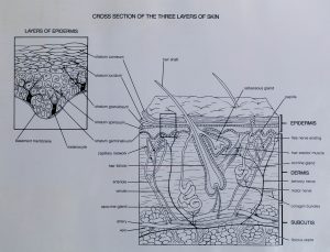
Image: “Cross Section of the Three Layers of Skin” by trustypics is licensed under CC BY-NC 2.0
Dermis
The second layer of skin is called the dermis. The dermis consists of fibrous and elastic tissue that forms a thick layer, giving the skin flexibility and strength. The dermis also contains nerve endings, sweat glands, sebaceous glands, hair follicles, and blood vessels. Different parts of the body will have varying numbers of these elements. For instance, your scalp has numerous hair follicles, but the palms of your hands and soles of your feet have none.
The primary function of the dermis is to help cushion/protect the body and to enable the skin to sense stimuli.
Subcutaneous layer
The subcutaneous layer is sometimes called the fat (adipose) layer or hypodermis layer of the skin. It is the innermost layer and consists of fat and connective tissues. Like the Dermis, the subcutaneous layer helps cushion the body and protect it from injury. However, this layer also has the important role of regulating body temperature. This layer varies in thickness depending on its position on the body. It is generally a fraction of an inch on your eyelids, but may be several inches thick on your abdomen and buttocks.
The subcutaneous layer also connects the skin to the muscles and bones through specialized connective tissue.
Glands
The skin contains two major types of glands: sweat glands and sebaceous glands.
Sweat glands
Sweat glands (sudoriferous glands) are located primarily in the subcutaneous layer of the skin and serve to help regulate body temperature. In addition to regulating temperature, sweat glands also help remove waste through the secretion of sweat.
Sweat glands are found throughout the body but are most numerous on your forehead, armpits, palms and soles of your feet.
Sebaceous gland
Sebaceous glands produce and secrete sebum which is an oily substance that coats and moisturizes your skin. This substance also has antibacterial and antifungal properties.
The production of sebum starts to diminish around the age of 20 and continues to decrease with age.
Hair and Nails
Two of the accessory structures of the Integumentary System are hair and nails.
Hair
As mentioned previously, hair is found on nearly every part of the human body. It is anchored to the skin through hair follicles, which are small, tube-like structures within the dermis layer of the skin. The growth of hair begins in a cluster of cells called papilla. As long as the papilla is viable, new hair will grow to replace any removed hair.
The primary purposes of hair are protection and body temperature regulation. When it is cold outside, the muscles surrounding the hair follicle cause the hair to stand up and trap more heat. When it is warm outside, your sweat glands produce sweat which is caught in the short hairs covering your body and helps cool you down.
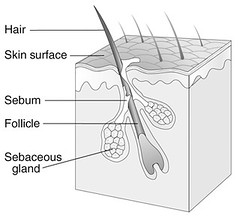
Image: “Normal Pilosebaceous Unit” by National Institute of Arthritis and Musculoskeleta is marked with CC PDM 1.0
The hair in our nose and eyelashes help keep foreign debris from entering. The hair in our nose also helps maintain moisture in the air we breathe. Our eyebrows and the hair on our heads provide protection from the sun. Eyebrows also help keep moisture from getting into our eyes.
Nails
The nails on our hands and feet are composed of keratin.
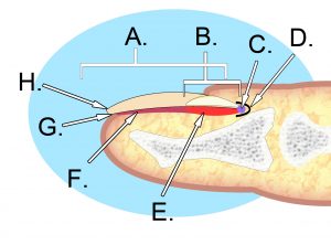
Image: “File:Human nail anatomy.jpg” by KDS444 is licensed under CC BY-SA 3.0
A: nail plate; B: lunula; C: root; D: sinus; E: matrix; F: nail bed: G: hyponychium; H: free margin
Nail plate
This is the hardest part of the nail and is made of hardened keratin. The layers of dead, compacted cells create a strong but flexible shape.
Lunula
This is the visible portion of the nail matrix. It is white and half-moon in shape.
Root
The nail root is embedded under the skin and attached to the matrix.
Sinus
This is where the nail root is located.
Matrix
This is the area where the nail starts to grow. The matrix creates new cells which push out the older cells and form nails.
Nail bed
The nailbed is composed of the skin (epidermis and dermis) beneath the nail plate.
Hyponychium
The hyponychium is the skin just beneath the free edge of the nail, near your fingertip. It is sometimes referred to as the “quick.”
Free margin
This is the end or distal portion of the nail plate. It is the portion of the nail that can be cut without damage.
Combining Forms Related to the Integumentary System
The following table includes combining forms related to the Integumentary System. It is not inclusive but does contain a fairly comprehensive list. This list also includes some related combining forms not specifically connected to the Integumentary System.
|
Combining Form |
Pronunciation |
Meaning |
Example |
|
aden/o |
ă-dĭn-ō |
gland |
adenocyst (a cystic tumor arising from a gland) |
|
adip/o lip/o steat/o |
ă-dĭp-ō lĭp-ō stē-ăt-ō |
fat |
adiposis (excessive accumulation of fat) lipocyte (fat cell) steatoma (fatty tumor) |
|
albin/o leuk/o |
ăl-bī-nō loo-kō |
white |
albinuria (pertaining to passing of white or colorless urine of low specific gravity) leukocyte (white blood cell) |
|
bas/i basi/o bas/o |
bās-ē bās-ē-ō bās-ō |
base |
basifacial (relating to the lower portion of the face) basophil (a cell with granules that stain with basic dyes) |
|
chromat/o |
krō-măt-ō |
color |
chromatic (pertaining to color) |
|
cirrh/o xanth/o |
sĭr-ō zăn-thō |
yellow |
cirrhosis (abnormal condition of the liver, causing a yellow tinge to the skin) xanthous (pertaining to yellow)
|
|
cutane/o derm/o dermat/o |
kū-tā-nē-ō dĕrm-ō dĕrm-ăt-ō |
skin |
cutaneous (pertaining to the skin) dermal (pertaining to the skin) dermatitis (inflammation of the skin) |
|
cyan/o |
sī-ăn-ō |
blue |
cyanosis (abnormal condition of bluish skin) |
|
cyt/o |
sīt-ō |
cell |
cytocide (an agent that kills cells) |
|
erythem/o erythemat/o erythr/o |
ĕr-ĭ-thē-mō ĕr-ĭ-thē-măt-ō ĕr-ĭth-rō |
red |
erythema (reddening of the skin) erythematous (pertaining to red) erythrocyte (red blood cell) |
|
eti/o |
ĕ-tē-ō |
cause, causation |
etiology (study of the causes of disease) |
|
hidr/o sudor/o |
hī-drō sūd-ōr-ō |
sweat |
hidrosis (abnormal condition of sweat; excessive sweating) sudoriferous (conveying or producing sweat) |
|
hydr/o |
hī-drō |
water |
hydrocele (the accumulation of serous fluid in a saclike cavity) |
|
(note: hidr/o and hydr/o are pronounced the same but have different meanings) |
|||
|
ichthy/o |
ĭk-thē-ō |
Fish, dry, scaly |
ichthyosis (abnormal condition in which the skin is dry and scaly, resembling fish skin) |
|
idi/o |
ĭd-ē-ō |
unknown, peculiar |
idiopathic (relating to a disease of unknown origin/cause) |
|
kerat/o |
kĕr-ă-tō |
horny, hard tissue; also means cornea |
keratinocyte (any of the cells in the skin that synthesize keratin) |
|
melan/o |
mĕl-ŭh-nō |
black |
melanoma (cancerous tumor of dark, blackish pigmented cells) |
|
morph/o |
mōr-fō |
form, shape |
morphometry (measurement of shapes/forms) |
|
myc/o |
mī-cō |
fungus |
mycoid (resembling fungus; fungus-like) |
|
necr/o |
nĕk-rō |
dead |
necrosis (death of cells, tissues or organs) |
|
onych/o |
ŏ-nĕ-kō |
nail |
onychomalacia (softening of the nail) |
|
path/o |
păth-ō |
disease |
pathology (the study of disease) |
|
pil/o trich/o |
pī-lō trĭk-ō |
hair |
pilonidal (pertaining to containing hairs; most often in dermoid cysts) trichoanesthesia (pertaining to loss of sensation of the hair) |
|
rhytid/o |
rĭ-tĭd-ō |
wrinkle |
rhytidectomy (surgical removal of wrinkles by plastic surgery) |
|
scler/o |
sklĕr-ō |
hardening; also means sclera |
sclerodermatitis (inflammation of the skin, accompanied by thickening and hardening) |
|
seb/o |
sĕb-ō |
sebum |
Seborrhea (overactivity of the sebaceous glands, resulting in an excessive amount of sebum) |
|
squam/o |
skwā-mō |
scale |
squamous (pertaining to scale-like) |
|
therm/o |
thĕr-mō |
heat |
thermometer (an instrument for measuring heat) |
|
xen/o |
zēn-ō |
foreign, strange |
xenogenic (pertaining to originating outside of the organism) |
|
xer/o |
zēr-ō |
dry |
xeroderma (dry skin) |
Abbreviations
The following is a list of abbreviations related to the Integumentary System, but is not intended to be all-inclusive.
|
Abbreviation |
Meaning |
|
Abbreviation |
Meaning |
|
BCC |
basal cell carcinoma |
|
ID |
intradermal (injection) |
|
Bx, bx |
biopsy |
|
IP |
ice pack |
|
C&S |
culture and sensitivity |
|
IV |
intravenous |
|
decub |
decubitus ulcer (also known as a pressure ulcer) |
|
MHP |
moist hot pack |
|
Derm. |
dermatology |
|
MM |
malignant melanoma |
|
Dx, dx |
diagnosis |
|
PE |
physical examination |
|
FH |
family history |
|
PPD |
purified protein derivative (skin test for tuberculosis) |
|
FS |
frozen section |
|
SCC |
squamous cell carcinoma |
|
H |
hypodermic |
|
SubQ, Sub-Q |
subcutaneous |
|
H&P |
history and physical |
|
Tx, tx |
treatment |
|
Hx, hx |
history |
|
ung. |
ointment |
|
I&D |
incision and drainage |
|
UV |
ultraviolet |
Common Tests and Procedures
The following is a list of common tests and procedures relating to the Integumentary System, but is not intended to be all-inclusive.
|
Test/procedure |
Explanation |
|
biopsy |
removal of a tissue sample for microscopic examination |
|
Botox injection |
injection used primarily to reduce facial wrinkles by preventing muscles from contracting; may also be used for back/neck spasms, overactive bladder, and migraines |
|
chemical peel |
applying a chemical solution to the skin, causing exfoliation |
|
debridement |
removal of dead/damaged tissue |
|
dermabrasion |
surgical removal of damaged or unwanted skin (moles, tattoos, scars, wrinkles) using abrasive materials |
|
dermatome |
an instrument for cutting thin slices of skin for grafting |
|
laser resurfacing |
Treatment used to reduce facial wrinkles and skin irregularities by directing short, concentrated pulsating beams of light, removing the treated skin layer by layer |
|
patch test |
suspected allergens are placed on the skin’s surface and covered for several days to see if/how the skin reacts |
|
punch biopsy |
type of biopsy that uses an instrument similar to a hole-punch to remove a sample |
|
scratch test |
Suspected allergens are scratched into the skin using an instrument to see if/how the skin reacts |
|
Wood’s lamp |
Produces an ultraviolet light used to diagnose various species of tinea manus (ringworm) and other bacterial or fungal infections |
Common Pathology Terms
Naturally, there are many types of diseases and conditions related to the Integumentary System – far too many to list in this text. However, the following list includes some of the most common.
|
Disease/condition |
Description |
|
Acne |
Inflammatory skin disorder often caused by overactive sebaceous glands. It can also be caused by genetics, hormones, menstruation, stress, foods with high glycemic (sugar) content, and some medications. |
|
Actinic keratosis |
Precancerous condition; scaly spots or patches on the top layer of the skin caused by chronic sun exposure |
|
Alopecia |
Condition causing hair to fall out in small patches when your autoimmune system attacks the hair follicles |
|
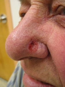
Basal cell carcinoma |
A common type of slow-growing skin cancer caused by chronic sun exposure. BCCs may look like open sores, red patches on the skin, or shiny bumps or growths.
Image: “File:Basal cell carcinoma2.JPG” by James Heilman, MD is licensed under CC BY 3.0 |
|
Burn |
Burns are thermal injuries to the skin caused by a variety of heat sources. Burns are typically classified as first-degree, second-degree, and third-degree. A first-degree burn will appear as red, non-blistered skin. A second-degree burn contains blisters with some thickening of the skin. A third-degree burn has a leathery appearance with a high degree of thickness. This is a healing, second-degree burn.
Image: “second degree burn healing (day 5)” by Mindfu
l One is licensed under CC BY-NC-ND 2.0
This is a severe, third-degree burn.
Image: “Advanced third degree burns” by Rosenwald is licensed under CC BY-SA 2.0
|
|
Callus |
Thickened, hardened part of the skin, commonly found in areas of the skin subject to frequent friction |
|
Carbuncle |
Cluster of red, swollen and painful boils connected to each other under the skin often caused by the bacteria Staphylococcus aureus |
|
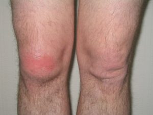
Cellulitis |
Painful, and sometimes serious bacterial skin infection, often affecting the lower legs. It often first appears as a red, swollen area that is hot and tender to touch. If untreated, it can become life threatening.
“Early cellulitis right knee” by jlcampbell104 is marked with CC PDM 1.0 |
|
Chloasma |
Brownish, irregularly shaped patches, often found on the faces of pregnant women |
|
Comedo |
Caused by a clogged hair follicle. It can be open (blackhead) or closed (whitehead) and occur with/without acne |
|
Contusion |
Bruise; discoloration of the skin after small veins and capillaries under the skin break |
|
Cyst (skin) |
Fluid-filled lump just under the skin |
|
Decubitus ulcer (pressure ulcer, bedsore) |
Open wound on the skin formed when the pressure from the body’s weight cuts off the blood supply to the skin and injures the tissues. Mostly commonly form on the buttocks/hips, back/shoulder blades, tailbone, ankles/heel, and elbows. There are four stages. In stage 1, the skin is red or discolored, but is still intact; in stage II, the skin breaks open to review a shallow ulcer or blister; in stage III, the ulcer is deeper and affects the subcutaneous/fat layer of the skin and the wound may contain pus; in stage IV, the ulcer moves deeper and affects the muscles and/or bone. |
|
Dermatitis |
Inflammation of the skin, caused by a variety of conditions. |
|
Ecchymosis |
A discoloration of the skin caused by bleeding underneath (bruise). |
|
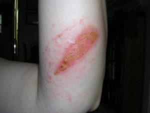
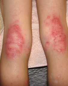
Eczema |
Condition that causes the skin to become red and itchy. It may be accompanied by asthma or hay fever.
Image: “Eczema behind knee” by Care_SMC is licensed under CC BY-ND 2.0 |
|
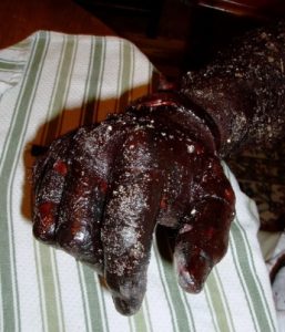
Frostbite |
Injury occuring when the skin is exposed to extremely cold conditions, causing the skin and underlying tissues to freeze. It is most common on the fingers, toes, nose, ears, cheeks and chin. In cases of extreme frostbite, the skin will turn black and hard as the tissue dies. |
|
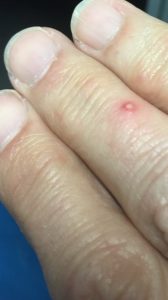
Furnuncle (boil) |
Infection of a hair follicle and nearby tissue |
|
Hematoma |
Bruise; An area of blood that has collected outside of the larger blood vessels, commonly due to injuries or trauma. Similar to a contusion, but involving larger blood vessels. |
|
Hirtsuism |
Excessive hair growth in women resulting in the growth of dark/coarse hair in a male-like pattern. |
|
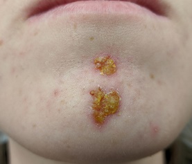
Impetigo |
Highly contagious skin infection that appears as red sores on the face around the nose and mouth or the hands and feet. When the sores burst, they develop a yellowish crust. Most commonly found in infants and children.
Image By James Heilman, MD – Own work, CC BY-SA 4.0,https://commons.wikimedia.org/w/index.php?curid=90030642 |
|
Laceration |
Cut or tear on the skin |
|
Lyme Disease |
Bacterial infection transmitted by infected ticks. Symptoms generally start with a rash, fever, headache and fatigue, but if untreated, may spread to the joints, heart, and nervous system.
Image: “File:Bullseye Lyme Disease Rash.jpg” by Hannah Garrison is licensed under CC BY-SA 2.5 |
|
Macule |
Small spots or blemishes on the surface of the skin. Freckles are a type of macule. |
|
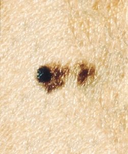
Malignant melanoma |
An aggressive form of skin cancer caused by exposure to ultraviolet light. It can develop anywhere on the body but most commonly occurs on areas that have had prolonged sun exposure, such as the back, legs, arms, and face. The first signs include: changes in an existing mole or thedevelopment of new pigmented or unusual-looking skin growths. To help identify MM, think of the letters ABCDE. A = assymmetrical shape; B = irregular border; C = changes in color or uneven distribution of color; D = diameter larger than ¼ inch; E = evolving or changing over time. MM may present with all of the signs or just one or two.
Image By Unknown photographer – This image was released by the National Cancer Institute, an agency part of the National Institutes of Health, with the ID 9188 (image) (next)., Public Domain, https://commons.wikimedia.org/w/index.php?curid=24053009 |
|
Papule |
Small, raised spot or bump on the skin |
|
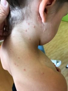
Pediculosis (lice) |
Infestation of hairy parts of the body, such as the head, body, or pubic region by lice, causing itching. Pediculosis is easily transferred from person to person during direct contact, including wearing of infected clothing. |
|
Petechiae |
Tiny purple, red, or brown spots on the skin, usually the arms, legs, stomach, and buttocks, caused by bleeding under the skin. When you press on the spots, they will not turn white. The cause of petechiae can be an infection or a reaction to medications. |
|
Psoriasis |
Chronic, inflammatory skin disorder marked by the developing of reddenned patches of skin with whitish, scaly plaques or patches, most commonly found on the knees, elbows, trunk, and scalp. The affected skin may itch, burn, and sometimes bleed. It is not contagious.
Image By Gzzz – Own work, CC BY-SA 4.0, https://commons.wikimedia.org/w/index.php?curid=88694219 |
|
Pustule |
Small bumps on the skin containing fluid or pus. They may develop on any part of the body, but are found most commonly on the back, chest and face.
“Pustule from Fire Ant bite” by euthman is licensed under CC BY 2.0 |
|
Rosacea |
Chronic condition that causes flushing and redness of the face, neck, and chest. Sometimes, small, red, pus-filled bumps also appear. Symptoms may come and go. |
|
Scabies |
An itchy skin condition caused by a tiny, burrowing mite. Scabies is extremely contagious and spread through close physical contact. Signs of scabies include itching and thin, irregular burrow tracks on the skin. |
|
Seborrheic keratosis |
Noncancerous skin growth, usually brown, black, or light tan that look waxy, scaly, and slightly raised. |
|
Squamous cell carcinoma |
Type of cancer commonly found in the mouth, esophagus, bronchi, lungs, vagina, and uterine cervix. It is the second most common form of skin cancer and, if caught early, is curable. SCCs may appear as scaly red patches, open sores, rough/thickened skin, or raised growths with a central depression. They may crust over, itch, or bleed. |
|
Tinea |
A group of fungal dieases including ringworm, arthlete’s food, and jock itch. |
|
Vesicle |
Clear, fluid-filled blister |
|
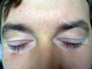
Vitiligo |
Chronic skin disease that results in patchy areas without pigmentation. There is no known cause.
Image: “File:Eyelid vitiligo 06.jpg” by Maria Sieglinda von Nudeldorf is licensed under CC BY-SA 4.0 |
|
Wart |
Small, benign skin tumor caused by the human papilomavirus (HPV). |
|
Wheal (welt, hive) |
A redish, swollen, itchy area on the skin, often a sign of an allergy. |

Practice Activities
Part of learning medical terminology is to practice defining terms based on their word elements. As discussed in Chapter 1, these definitions will provide a general definition, but may need to be refined using a medical dictionary.
If you remember, when you define a medical term based on the word elements, you will:
- Define the suffix or last part of the word first
- Define the first part of the word next (which may be a prefix or a word root)
- Then define the remaining word parts in order.
Adipoid contains the following word parts: adip/o (meaning fat) and –oid (suffix meaning resembling). Therefore based on the word parts, adipoid would be defined as “resembling fat.” In Taber’s 20th edition, this word is defined as “Fatlike; lipoid.”
The word idiopathic contains 3 word elements: idi/o (meaning unknown or peculiar), path/o (meaning disease), and –ic (suffix meaning pertaining to). Based on the word parts, idiopathic would be defined as “pertaining to unknown or peculiar disease.” Taber’s 20th edition defines iodiopathic as “pertaining to illnesses whose cause is either uncertain or as yet undetermined.”
Your turn
Activity 1
To assist in your learning, break the following medical terms into their word elements and define the medical term based on the meaning of each word part. Then, using a medical dictionary of your choice, compare the word part definition to the dictionary definition.
- Cyanotic
- Hidradenitis
- Xerocyte
- Dermatomycosis
- Pathologist
- Hydrotherapy
- Hypodermic
- Sclerodermatitis
- Onychomycosis
- Dermoplasty
Activity 2
Create medical terms to match the following definitions.
- Inflammation of the skin
- Tumor of the nail
- Disease of the hair
- Surgical excision of fat
- Abnormal condition of no sweat
- Pertaining to redness
- Study of cells
- Abnormal condition of blue
- Pertaining to color
- The study of shapes or forms
Activity 1 Answers
Your dictionary definition will vary depending on the one you used, but the word parts are defined as follows:
- Cyanotic: cyan/o + -tic (pertaining to blue)
- Hidradenitis: hidr/o + aden/o + -it is (inflammation of sweat gland)
- Xerocyte: xer/o + -cyte (cell dry; Note: this is a medical term that doesn’t follow the normal definition guidelines. It would make more sense to define it as a “dry cell.”)
- Dermatomycosis: dermat/o + myc/o + -osis (abnormal condition of skin fungus)
- Pathologist: path/o + -ologist (specialist in the study of disease)
- Hydrotherapy: hydr/o + -therapy (treatment [using] water)
- Hypodermic: hypo- + derm/o + -ic (pertaining to below the skin)
- Sclerodermatitis: scler/o + dermat/o + -itis (inflammation [causing] hardening of the skin
- Onychomycosis: onych/o + myc/o + -osis (abnormal condition of nail fungus)
- Dermoplasty: derm/o + -plasty (surgical repair of the skin)
Activity 2 Answers
Although several of the definitions have multiple combining forms and/or suffixes that mean the same thing, the answers below reflect the most common version of the term.
- Dermatitis
- Onychoma
- Trichopathy
- Lipectomy
- Anhidrosis
- Erythematous
- Cytology
- Cyanosis
- Chromatic
- Morphology