
4 Chapter 4 – Sex Differentiation, Anatomy, and Physiology
Ericka Goerling, PhD and Emerson Wolfe, MS
Chapter 4 Learning Outcomes
- Distinguish between female, male, and intersex anatomy and recognize the differences and similarities between them
- Identify hormones that influence sex development and sexual behavior
- Explain how bipotential tissues are directed to develop into male or female sex organs
- Name the rudimentary duct systems in the embryo that are precursors to male or female internal sex organs
- Describe the hormonal changes that bring about puberty, and the secondary sex characteristics of men and women
- Analyze the complexity of sex development and gain an understanding of the experiences of intersex and transgender individuals
Every body is different. But many things about our bodies are the same.
Humans come in a variety of shades, sizes and proportions, yet in total, our bodies are more similar to each other than they are different. In fact the human body shares similarities with bodies across the diversity of life. We share aspects of our reproductive system with all mammals, aspects of our basic physiology with all vertebrates, and aspects of our cell structure, biochemistry and genetics with all living things. In this chapter, we will look at the human body specifically, not because the human body, in terms of reproduction, is very distinct from that of a 3-toed sloth, or because the basic structure is very different from a Galápagos tortoise, or because our cellular biology varies much from that of the fungus that inhabits bleu cheese. Instead, we focus on the human body because the authors and readers of this text presumably each have a human body, and reading about ourselves is interesting.
AUTHOR’S NOTE: Some of the language in this chapter is still held in the binary; that is girl/boy or male/female. This is concurrent with a lot of medical literature. Importantly, however, even biological sex is found on a continuum so there are variations of genital structure and presentation that defies the binary, which is critical to note.
Sex Differentiation
Sex differentiation, also referred to as sexual differentiation, is the process in which genitals and reproductive organs develop within the womb as a result of complex hormonal processes altering neutral tissues to develop along female and male lines and in which varying combinations are possible as well. Secondary sex characteristics begin to develop further as part of the sex differentiation process during puberty. Therefore, prenatal development, as well as changes that further occur during puberty, create a cascade of events that results in physical changes to the body. Chromosomes, genes, gonads, hormones, and hormone receptor sites play key roles within the endocrine system that influence sex development. Sex is both a genetic and environmental experience in which epigenetic factors can alter the way that genes function, resulting in changes to hormone production and hormone receptor sites at different points across the lifespan.
Prenatal
Commonalities between male and female reproductive anatomy
During embryonic development the male and female fetus are indistinguishable before about 10 weeks of pregnancy. Fetal tissues begin in an undifferentiated state, and based on genetic signals and the interuterine environment the reproductive organs usually differentiate into structures typical of males and females. This means that for most of the reproductive parts there is an analogous part in the other sex that arose from the same original tissues. For example, testes and ovaries develop from the same tissue – originally located in the abdomen. In males the testes move down and outside the abdomen as they develop; in female they remain internal. Some structures (such as the oviducts) have a structure that was common in early development, but completely or partially disappears in later development; other structures (such as the uterus) have analogues that are very subtle structures in the male. See the following table for a list of analogous structures in male and female anatomy.
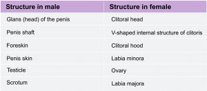
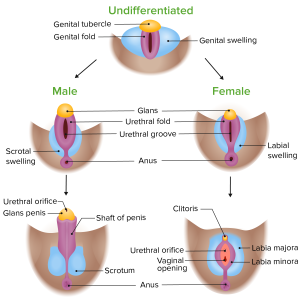
Image by Lecturio. License: CC BY-NC-SA 4.0
Commonalities between male and female reproductive signaling
Much of the reproductive physiology we will address is regulated by hormonal signals that arise in the brain and much of this signaling is shared between males and females.
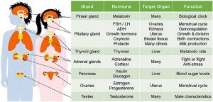
Within the brain is a region called the hypothalamus (see figure below). This portion of the brain sends signals to the pituitary gland located beneath it. In particular, the hypothalamus sends a hormonal signal called gonadotropin-releasing hormone (GRH) to the pituitary gland. In response to the GRH signal, the pituitary gland releases two hormones that circulate in the blood: luteinizing hormone (LH) and follicle stimulating hormone (FSH). These hormones travel throughout the body, triggering further hormone releases and physiological changes (discussed further below). There are feedback loops that tightly regulate the levels of circulating hormones. In addition to GRH, LH, and FSH, the hormones testosterone, estrogen and progesterone are important in reproductive signaling. While we will focus on the effects of testosterone in males and estrogen and progesterone in females, all of these hormones are present and important in both males and females.
Further Sexual Development Occurs at Puberty
Puberty is the stage of development at which individuals become sexually mature. Though the outcomes of puberty for boys and girls are very different, the hormonal control of the process is very similar. In addition, though the timing of these events varies between individuals, the sequence of changes that occur is predictable for male and female adolescents. As shown in the figure below, a concerted release of hormones from the hypothalamus (GnRH), the anterior pituitary (LH and FSH), and the gonads (either testosterone or estrogen) is responsible for the maturation of the reproductive systems and the development of secondary sex characteristics, which are physical changes that serve auxiliary roles in reproduction.
The first changes begin around the age of eight or nine when the production of LH becomes detectable. The release of LH occurs primarily at night during sleep and precedes the physical changes of puberty by several years. In pre-pubertal children, the sensitivity of the negative feedback system in the hypothalamus and pituitary is very high. This means that very low concentrations of androgens or estrogens will negatively feed back onto the hypothalamus and pituitary, keeping the production of GnRH, LH, and FSH low.
As an individual approaches puberty, two changes in sensitivity occur. The first is a decrease of sensitivity in the hypothalamus and pituitary to negative feedback, meaning that it takes increasingly larger concentrations of sex steroid hormones to stop the production of LH and FSH. The second change in sensitivity is an increase in sensitivity of the gonads to the FSH and LH signals, meaning the gonads of adults are more responsive to gonadotropins than are the gonads of children. As a result of these two changes, the levels of LH and FSH slowly increase and lead to the enlargement and maturation of the gonads, which in turn leads to secretion of higher levels of sex hormones and the initiation of spermatogenesis and folliculogenesis.
In addition to age, multiple factors can affect the age of onset of puberty, including genetics, environment, and psychological stress. One of the more important influences may be nutrition; historical data demonstrate the effect of better and more consistent nutrition on the age of menarche in girls in the United States, which decreased from an average age of approximately 17 years of age in 1860 to the current age of approximately 12.75 years in 1960, as it remains today. Some studies indicate a link between puberty onset and the amount of stored fat in an individual. This effect is more pronounced in girls, but has been documented in boys as well. Body fat, corresponding with secretion of the hormone leptin by adipose cells, appears to have a strong role in determining menarche. This may reflect to some extent the high metabolic costs of gestation and lactation. In girls who are lean and highly active, such as gymnasts, there is often a delay in the onset of puberty.
Kabotyanski and Somerville under the terms of the Creative Commons Attribution License (CC BY).
Hormones of Puberty During puberty, the release of LH and FSH from the anterior pituitary stimulates the gonads to produce sex hormones in both male and female adolescents.
Signs of Puberty
Different sex steroid hormone concentrations between the sexes in general and within each individual uniquely also contribute to the development and function of secondary sexual characteristics. Examples of secondary sexual characteristics are listed in Table 1. Each individual’s hormone concentrations will depend upon genetics, diet, stress, and more; thus, secondary sex characteristics will progress along a continuum of possibilities based on these nature multiplied by nurture combinations.
Development of the Secondary Sexual Characteristics
| Male | Female |
| Increased larynx size and deepening of the voice | Deposition of fat, predominantly in breasts and hips |
| Increased muscular development | Breast development |
| Growth of facial, axillary, and pubic hair, and increased growth of body hair | Broadening of the pelvis and growth of axillary, pubic and body hair |
Something to keep in mind as well is that people can experience varying degrees of muscular, breast and hair development depending upon their genetic and environmental makeup. For instance, some boys at the beginning of puberty may see breast tissue growth as well as other body fat increases, and some girls may have facial and body hair growth. One example in this complex process is how unused testosterone is converted to estrogen, for instance, making it not about how much testosterone is present and rather how that testosterone is being used or unused. Also, hormone receptor sites in female cells are often more reactive to lower levels of testosterone. Intersex individuals, females and males all have estrogen and testosterone impacting the changing body during puberty, and it is incorrect to view testosterone as a strictly “male” hormone and estrogen as a strictly “female” hormone. To understand this process, individuals will need to analyze their specific hormone levels, the functionality of genes involved in sex differentiation, and environmental stressors and nutrition that may have epigenetic effects on their genes.
As a girl reaches puberty, typically the first change that is visible is the development of the breast tissue. This is followed by the growth of axillary and pubic hair. A growth spurt normally starts at approximately age 9 to 11, and may last two years or more. During this time, a girl’s height can increase 3 inches a year. The next step in puberty is menarche, the start of menstruation.
In boys, the growth of the testes is typically the first physical sign of the beginning of puberty, which is followed by growth and pigmentation of the scrotum and growth of the penis. The next step is the growth of hair, including armpit, pubic, chest, and facial hair. Testosterone stimulates the growth of the larynx and thickening and lengthening of the vocal folds, which causes the voice to drop in pitch. The first fertile ejaculations typically appear at approximately 15 years of age, but this age can vary widely across individual boys. Unlike the early growth spurt observed in females, the male growth spurt occurs toward the end of puberty, at approximately age 11 to 13, and a boy’s height can increase as much as 4 inches a year. In some males, pubertal development can continue through the early 20s.
Hormone blockers for transgender individuals are helpful during puberty to prevent the development of inappropriate secondary sex characteristics that may evoke dysphoria. A concern around hormone blockers is that the parent(s) or guardian(s) are consenting to this treatment on their youth’s behalf, and some caretakers may not feel comfortable with this process. An ongoing debate exists around whether adolescents are mature and aware enough to advocate for themselves. Pediatricians and advocates who are supportive of prescribing hormone blockers and starting hormone replacement therapy express that transgender identities are often expressed during early childhood and remain consistent across the lifespan, whereas others who are against this process tend to express concern over the weight of this decision due to the epigenetic effects of hormone medications that will permanently alter the body and cannot be undone if an individual were to no longer identify as transgender.
Chromosomes and Hormones, Oh My!
Watch these two videos to gain more insight into the X chromosome and hormones.
And
Epigenetics, Surgery, and Medications
Epigenetics
Epigenetics is the way in which environmental factors influence the expression of genes. First, let’s look at reptilian and mammalian studies to explore some epigenetic factors that can influence differences between sexes. According to Forger (2016), “In many reptiles and some fish the ambient temperature during incubation determines the sex of the individual. In other words, perfectly good male or female brains and bodies can develop from an identical genome, based on differences in the epigenetic regulation of that genome. Similarly, although there are genetic differences between male and female mammals, many of the sex differences in mammalian brains and behavior are likely epigenetic in origin” (p. 1). In studies conducted on rodents, such as mice, rats, and guinea pigs who share similarities with human males regarding olfactory cues and some sexual behaviors, researchers have found that the development of the brain is influenced by environmental factors at birth that alter cell death in the brain and the way that sex hormones are processed based on activation or inactivation of key genes depending on the presence of certain histones (Forger, 2016).
Surgeries
The removal of organs, such as gonads, can alter the production of hormones and require an individual to take synthetically produced hormones. Hysterectomy (surgical removal of the uterus), orchiectomy (surgical removal of one or both testes), and other procedures that alter the reproductive organs or other endocrine glands will alter the natural production of hormones and cause changes to the body. Medications can be prescribed to stand in for the lost hormones that can no longer be created, but medications will require adjusting and tweaking over time which may never completely compare to the original levels previously maintained.
Medications
Many people associate medically prescribed hormones with transgender individuals who are physically transitioning; however, hormone replacement therapy was first developed to help cisgender women with symptoms related to menopause (Cagnacci & Venier, 2019). Additionally, testosterone injections and pills are often advertised to cisgender men under the premise that they will provide them with a confidence boost, higher sex drive, more muscle mass, etc. (Harvard Health Publishing, 2020). Hormones are present in birth control pills, and narcotics and prescribed substances, such as steroids, antihypertensive drugs, chemotherapy and more, can impact the endocrine system in numerous ways which will then impact the way that sex hormones are produced and processed. Thus, these medications beyond hormone replacement therapies can have impacts upon sexual functioning, fat distribution, mood, body hair production and more. Some changes, such as body hair production, may remain even after the medication is stopped.
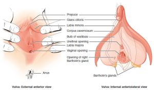
Female Anatomy
Some of the organs involved in female reproduction are diagrammed in the Figure below.
The reproductive structures of the human female are shown.
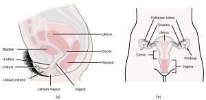
(credit a: modification of work by Gray’s Anatomy; credit b: modification of work by CDC)
Female reproductive anatomy includes external structures (the clitoris and vulva), structures involved in the production of eggs and in fetal development (ovaries corpus luteum, and uterus), and structures involved in the transport of sperm, eggs, and babies (vagina, cervix and oviduct).
For a useful 3-D interactive, check out Pussypedia’s 3-D Interactive Pussy.*

Pussy*
*We propose a new gender-and-organ-inclusive use of the word. ~Pussypedia
Egg production and fetal development
- Ovary: females have two ovaries that are the site of egg production, and, if an egg is fertilized, the site of the corpus luteum. The ovary produces hormones estrogen and progesterone and testosterone.
- Corpus luteum: the site of egg maturation within the ovary. After ovulation (release of the egg) the corpus luteum produces progesterone to maintain a possible pregnancy.
- Uterus: this muscle-lined, triangular organ is where a fertilized egg implants and develops. This organ develops a thick blood lining and sheds this lining on a monthly cycle.
Transport of eggs, sperm, and embryos
- Vagina: a highly expandable pouch structure that serves as the opening of the female reproductive tract to outside the body. The vagina is the point of sperm entry, and the point of exit for unfertilized eggs, menstrual discharge and for babies, if pregnant and having a vaginal delivery.
- Cervix: the opening between the vagina and the uterus. The size of this opening varies from tightly closed – to open for the passage of sperm, to open enough for a baby to pass through.
- Oviducts (sometimes called fallopian tubes): these ducts transport mature eggs from the ovary toward the uterus. If a sperm and egg are in the oviduct at the same time, the egg can be fertilized by a sperm.
Exterior structures
- Vulva: a general term for the exterior parts surrounding the vagina, including the labia majora and labia minora, which are the folds of skin on either side of the clitoris, urethra, and vagina. Often this term is overlooked, with folks referring to the vulva as “vagina,” which is the internal structure. It’s OK and more accurate to say vulva when referring to the external structure. Importantly, the structure and appearance of the vulva may vary widely. There’s no “one size fits all” when it comes to the vulva and diversity in appearance needs to be celebrated.
- Clitoris: the sensitive nerve-rich organ that is analogous to the head of the penis. The part of the clitoris that is visible outside the body is dorsal to (closer to the belly) the urethra and the vagina). The interior part of the clitoris extends internally along either side of the vagina.
Unfortunately, the structure of the clitoris is not well known by many people, including to those folks with a clitoris. Historically (and even in some contemporary settings), this feature of anatomy has been muted or correlated with antiquated notions of female sexuality such as hysteria or neurosis.
Female reproductive physiology
Female reproductive anatomy and physiology has many similarities to that of the male. As described earlier, females also use LH and FSH secretion from the pituitary triggered by GRH from the hypothalamus to stimulate hormone production by the gonads. There is also negative feedback to regulate hormone production. However, in females, the interplay among the hormonal signals is more complicated than in males. While male hormonal feedbacks and signaling provide a relatively steady level of the sex hormone testosterone, for females there is a monthly cycle over which the circulating hormone levels go up and down at the same time as changes occur in the ovaries and the uterus. This surging of hormones along with the changes in the ovaries and uterus require the more complicated physiological controls described below.
Menstruation
Sometimes a Taboo Topic
The banner in the picture below was carried in a 2014 march in Uganda as part of the celebration of Menstrual Hygiene Day. Menstrual Hygiene Day is an awareness day on May 28 of each year that aims to raise awareness worldwide about menstruation and menstrual hygiene. Maintaining good menstrual hygiene is difficult in developing countries like Uganda because of taboos on discussing menstruation and lack of availability of menstrual hygiene products. Poor menstrual hygiene, in turn, can lead to embarrassment, degradation, and reproductive health problems in females. May 28 was chosen as Menstrual Hygiene Day because of its symbolism. May is the fifth month of the year, and most women average five days of menstrual bleeding each month. The 28th day was chosen because the menstrual cycle averages about 28 days.

World Menstrual Hygiene Day celebration by Satirtha- The Helping Hand a non profit organization.
The monthly female reproductive cycle can be divided into three phases, the follicular phase, ovulation, and the luteal phase. For each of these phases, there are concurrent changes happening in the uterus and in the ovaries. See the figure below for a diagram of the phases of the female reproductive cycle and what is happening in the ovary, the uterus, and circulating hormone levels.
Female hormone cycle.
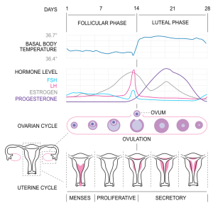
CC BY-SA 3.0
The Follicular Phase
The name “follicular phase” is in reference to the egg-containing follicle in the ovary that matures during this phase (note the ovarian histology shown in Figure 7). This phase begins on day one of a female’s reproductive cycle. Day one is defined as the first day of menstruation (the first day of a period). Menstruation occurs for about the first 5 days of the follicular phase. During these days, if a female is not pregnant, low circulating levels of the hormone progesterone trigger the breakdown of the endometrium (the lining of the uterus). This blood-rich tissue exits the uterus through the cervix and then leaves the body out the vagina. During menstruation, low circulating levels of estrogen and progesterone stimulate GRH production (from the hypothalamus in the brain), which leads to LH and FSH secretion by the pituitary gland. FSH signals the maturation of several follicles within the ovaries. These follicle cells produce a steadily increasing amount of estrogen (note the estradiol (a type of estrogen) increases from day 1-12 in Figure 4.5).
Ovulation
Ovulation refers to the rupture of a mature follicle within the ovary; this ruptured follicle releases an oocyte (an unfertilized egg) into the abdominal space. Because this rupture is an actual breakage, some females will feel a twinge or slight pain during ovulation. Ovulation generally happens around day 14 of the reproductive cycle in one ovary.
Luteal phase
During the luteal phase, the now empty follicle within the ovary collapses. This collapsed mass of cells is called a corpus luteum. The corpus luteum produces progesterone that enters the blood circulation. Progesterone signals the hypothalamus to signal the pituitary to reduce FSH and LH production, which prevents other follicles from maturing. If the oocyte in the oviduct is not fertilized, the corpus luteum degrades, causing a drop in progesterone, which triggers the beginning of menstruation and the return to the follicular phase of the reproductive cycle (back to day 1 after about a 28-day cycle). If the oocyte IS fertilized, then that begins the cellular process of fetal development (pregnancy).
Menopause
Female fertility (the ability to conceive) peaks when women are in their twenties, and is slowly reduced until a women reaches 35 years of age. After that time, fertility declines more rapidly, until it ends completely at the end of menopause. Menopause is the cessation of the menstrual cycle that occurs as a result of the loss of ovarian follicles and the hormones that they produce. A woman is considered to have completed menopause if she has not menstruated in a full year. After that point, she is considered postmenopausal. The average age for this change is consistent worldwide at between 50 and 52 years of age, but it can normally occur in a woman’s forties, or later in her fifties. Poor health, including smoking, can lead to earlier loss of fertility and earlier menopause.
Intersex Anatomy
About 1 in 1,000-1,500 people will be born noticeably intersex, such as having partial elements of both a penis and vulva. However, other intersex conditions may not show up until later, such as during puberty or when trying to conceive children, making this number higher in actuality. Some estimates show that some intersex conditions can be as high as 1 in 66. Taken altogether, some researchers, such as Anne Fausto-Sterling, argue that the number of intersex people is actually closer to about 1 in 100 (Intersex Society of North America, 2008b). Here is a video of 4 individuals sharing some of their experiences:
The term intersex is an umbrella term that encompasses many different variations to sex. Some intersex individuals have variations to their sex chromosomes or that the SRY gene on the Y chromosome is not present or was translocated onto an X chromosome. Thus, in some instances, a male can have XX chromosomes and a female can have XY chromosomes. Other variations are possible as well (X0, XXY, XYY, XXX). According to Dr. Charmian Quigley with the Intersex Society of North America (2008a), “The last time I counted, there were at least 30 genes that have been found to have important roles in the development of sex in either humans or mice. Of these 30 or so genes 3 are located on the X chromosome, 1 on the Y chromosome and the rest are on other chromosomes, called autosomes (on chromosomes 1, 2, 3, 4, 7, 8, 9, 10, 11, 12, 17, 19). In light of this, sex should be considered not a product of our chromosomes, but rather, a product of our total genetic makeup, and of the functions of these genes during development” (para. 11-12). Therefore, we need to be careful when we oversimplify this process and relate sex to XX or XY chromosomes only.
Male Anatomy
Some of the organs involved with male reproduction are diagrammed below.

Male reproductive anatomy
Male reproductive anatomy involves the organs and glands that produce sperm, create semen to transport sperm, and conduct this liquid semen out of the body. Semen production involves the work of accessory glands, each responsible for the production of one or more key ingredients of semen. Male anatomical structures can be broken down into the following:
Sperm production
- Testis: males typically have two testes (also called testicles), which, in humans, descend from the abdomen during fetal development and are enclosed outside the abdomen in the scrotum. Each testis houses many tube-like structures (the seminiferous tubules) in which sperm are made. Specialized cells (the Leydig cells) in the testes produce testosterone.
- Scrotum: a pouch of skin that holds the testes that contracts or expands to adjust the distance the testes are from the body to regulate their temperature.
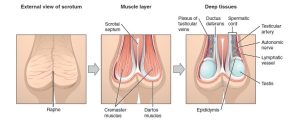
The central raphe on the far left represents the “seam” where the genital tubercles fused in development. In the central image, note the cremaster and dartos muscles, which are important for temperature regulation. On the right, note a deep layer demonstrating the external testes, epididymis, and neurovasculature.
Image: “The Scrotum and Testes” by Phil Schatz. License: CC BY 4.0
- Seminiferous tubules: these structures within the testes are the actual sites of sperm production (discussed further below)
- Epididymis: this rubbery device sits astride the testis. Sperm mature here and are stored prior to ejaculation (when sperm-bearing semen leaves the body, typically during orgasm)
Semen production
- Seminal vesicles: these two glands produce an alkaline (basic) fluid that can neutralize the acidity of the vagina. This fluid contains fructose and other nutrients to provide energy for the sperm.

- Bulbourethral (or Cowper’s) glands: these two glands provide a mucus-rich alkaline fluid that lubricates the inside of the urethra to allow for easier passage of sperm and neutralizes the urethra (urine residue is acidic). Some of this fluid exits the penis prior to ejaculation (this pre-ejaculate fluid can also contain sperm). The remainder of the fluid combines with the semen ejaculate.
- Prostate gland: this organ wraps around the urethra and provides muscular contractions that help propel semen during ejaculation and block urine flow from the bladder during ejaculation. It also provides fluid in the ejaculate that contains enzymes and zinc that aid in sperm motility.
Sperm/semen transport:
- Ductus (or vas) deferens: this pair of muscle-lined tubes carry sperm from the epididymis of each testis into the abdominal cavity where they loop over the bladder and join with the ducts from the seminal vesicles to form the ejaculatory ducts. The muscles that line the ductus deferens contract to propel semen during ejaculation.
- Ejaculatory ducts: these ducts are formed by the joining of the vas deference with the duct from the seminal vesicle. Each ejaculatory duct empties into the urethra.
- Penis: the organ that encircles the urethra as the urethra exits the abdomen. This organ changes from flaccid (soft and limp) to erect (rigid and standing away from the body) during sexual arousal or spontaneously. In uncircumcised men the penis has a fold of skin called a foreskin that during the flaccid state, covers the head of the penis, and during the erect state retracts behind the glans (or head) of the penis.
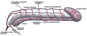
Image: “Arterial supply of the penis” by Gray’s anatomy. License: Public Domain
- Urethra: the tube that runs from the bladder through the penis through which urine and semen exit the body.
Transgender Anatomy
Further neurobiological research has been conducted to explain why many transgender people experience gender dysphoria, which is an intense feeling that one’s sex assigned at birth based on genital presentation does not match the way they feel about themselves. The contrast between the genital-based presentation of sex and the brain’s sex can be explained from a neurobiology perspective by taking a closer look at sex differentiation when we are still in the womb. During prenatal development, sex differentiation of the genitals takes place during the first two months of pregnancy while sex differentiation of the brain occurs around 4 months of pregnancy (Swaab & Garcia-Falgueras, 2009). This discrepancy between the development of the genitals and the brain has led biological researchers to believe this may be the reason why some individuals are transgender or experience gender dysphoria. Something to keep in mind: Humans actually show a lot less sex dimorphism than many other species, which means that there are actually a lot of similarities between men and women, making this type of research rather difficult as differences are often very subtle if present at all.
Due to the positive results found with treating some transgender individuals using hormone replacement therapy and hormone blockers in order to bring about desirable changes to the physical body, this adds merit to the perspective that there may be some biological reasons and solutions for people experiencing gender dysphoria from a biopsycho perspective. Transgender men: testosterone therapy will result in enlargement of the clitoris leading to resembling a small penis, facial hair and increased body hair similar to other males in the family, redistribution of body fat and increased muscle mass, deepening of the voice due to testosterone thickening the vocal cords, breast tissue will begin to atrophy, more oily skin, etc. Transgender women: Hormone blockers and estrogen hormone therapy will cause breast tissue growth, redistribution of body fat and reduced muscle mass, body hair will thin and grow less fast, penial tissue (if not having regular sex or masturbating) will begin to atrophy, the skin will become drier, etc. Electrolysis for hair removal may be desired and voice lessons can help train the individual to speak in a more socially expected female register.
Hormone blockers prior to puberty are especially beneficial to prevent these long-lasting impacts and aid the transition process for transgender individuals. Many parents feel conflicted about this, however, and may not consent on their children’s behalf to receive these medical services. Surgeries may also be used by some transgender individuals, commonly referred to within these communities as “top surgery” relating to surgery to either remove or enhance breast tissue or “bottom surgery” on the genitals. Creating a vagina can be done by utilizing the existing tissue and has promising results for many individuals allowing them to experience sensation, but this will depend on scar tissue. Creating a penis will require the addition of testicular implants and a penial pump for erection purposes.
Every transgender person will have to decide what steps they want to take to help them feel as comfortable as possible in their bodies. If someone does not use hormones or have surgeries, it does not make them any less transgender than someone who does. Also, keep in mind that access to healthcare is a privilege and not a right in our country, which can be a barrier for many individuals in receiving the care they need. Family, culture, society, etc. also act as barriers in this process. It is never appropriate to question a transgender person about their genitals, surgeries or hormones, and doing so would be examples of microaggressions. This is othering and dehumanizing and reduces transgender individuals to their genitals and body rather than seeing them as a whole person with feelings and the right to privacy. Create a space where someone feels comfortable to share their experiences with you on their own instead rather than questioning them. We will talk more about allyship next week. Until then, here are some resources:
- National Center for Transgender Equity
- Trans Student Educational Research
- Supporting the Transgender People in Your Life: A Guide to Being a Good Ally
Additional resources for your reference: If you or someone you know is interested in learning more about how hormone replacement therapy impacts the body, check out the links provided by the University of California San Francisco Transgender Care. UCSF Transgender Care also has more information on the types of surgeries that are available for transgender individuals.
What about when nonbinary individuals, such as those who identify as agender, bigender, genderqueer, etc., and experience gender dysphoria as well? Or, what about when transgender individuals don’t experience gender dysphoria? What might be some biological explanations? We need more research! A question to think about further: If gender is a social construct, then does biology even really matter anyway and are researchers trying too hard to find biological answers? This goes back to the nature/nurture debate once again. We will discuss gender as a social and cultural phenomenon next week to explore this question further from other perspectives.
To hear personal stories and experiences of transgender individuals, watch this video:
Bodies are Beautiful and Vary So Much
The authors of this text (Ericka Goerling and Emerson Wolfe) could not find images that represent transgender bodies, apart from images depicting surgeries, in a respectful way. If you know a transgender, non-binary, or gender expansive artist who would like to see their work featuring beautiful bodies added to this resource, please share your instructor’s email with them to reach out for more information on this project.
The Medicalization of Gender: The Development of “Sex”
Why do we label different anatomical parts as “female anatomy” and “male anatomy”? Our understanding of the body is usually through the lens of long deceased researchers and doctors who believed in strict gender binary systems with males having “normal” bodies and with women being framed as having less capable bodies. The existence of intersex people indicates that the process of sex differentiation is very complex, yet our understanding today even is misguided by an oversimplification of this process that is shaped by our cultural definition of gender and our gender schema (aka a cognitive blueprint based on your experiences). Think to your own upraising and explore how this has shaped your concept of sex and gender even today. We will discuss cross-cultural perspectives on gender and how this impacts our idea of biological sex next week. Until then, explore the following questions: Do you know your hormone levels? Do you know your sex chromosomes? Take this moment to normalize the way your genitals looked at birth and how they look currently and recognize that everyone’s bodies are different and beautiful.
Conclusion
As you reach the end of this reading, the hope is that you have more questions than answers about the vast possibilities that contribute to your present bodily form and physiological functioning. Human experiences are genetic and environmental, and sex differentiation further showcases how this process is often oversimplified and discussed from within binary systems of “female” and “male” when the reality lends itself more toward spectrums and continuums from the moment we begin in the womb to how we undergo many changes throughout our life’s course. Use this knowledge to understand your body’s functioning, and harness this information as you explore what brings you and others sexual pleasure if this connection is something you seek. Use scientific advancements to be allies to the human experience and also recognize the limitations to scientific study. We do not have all the answers and may never, so continue to ask questions about human sexual development, anatomy, and physiology.
Reflection Questions
- Sex differentiation is a complex process influenced by chromosomes, hormones, and environmental factors. How might this understanding challenge common assumptions about biological sex as a simple binary? What implications could this have for how we think about gender and sex?
- Consider the section on intersex conditions. How might the existence of intersex individuals impact our understanding of sex and gender? What ethical considerations arise when thinking about medical interventions for intersex infants?
- We note epigenetic factors that can influence sex differentiation. How does this information contribute to the nature vs. nurture debate regarding human sexuality and development? Can you think of any other areas where epigenetics might play a role in sexual development?
- Reflect on the information provided about transgender anatomy and physiology. How might this knowledge inform healthcare practices and policies regarding transgender individuals? What challenges might arise in providing comprehensive care for transgender patients?
- We conclude by questioning the medicalization of gender and the development of “sex” as a concept. How do you think historical and cultural factors have shaped our current understanding of sex and anatomy? How might this understanding evolve in the future as we learn more about human diversity?
Licenses and Attributions
Betts, J. G., Young, K. A., Wise, J. A., Johnson, E., Poe, B., Kruse, D. H., Korol, O., Johnson, J. E., Womble, M., & DeSaix, P. (2013). Anatomy and physiology. OpenStax. https://openstax.org/books/anatomy-and-physiology/pages/27-3-development-of-the-male-and-female-reproductive-systems#fig-ch28_03_01 (Adapted for the section on Puberty). Licensed under a Creative Commons Attribution-NonCommercial-ShareAlike 4.0 International License.
Cotner, S. & Wassenberg, D. (2018). The evolution and biology of sex. PressBooks.https://open.lib.umn.edu/evolutionbiology/ (for sections on Female Anatomy and Male Anatomy). Licensed under a Creative Commons Attribution-NonCommercial-ShareAlike 4.0 International License.
Miller, C. (2016). Human biology. PressBooks. https://humanbiology.pressbooks.tru.ca/. Licensed under a Creative Commons Attribution-NonCommercial-ShareAlike 4.0 International License.
OpenStax College. (2013). Anatomy & physiology. OpenStax. http://cnx.org/content/col11496/latest/. Licensed under a Creative Commons Attribution-NonCommercial-ShareAlike 4.0 International License.
Adaptations: Reformatted. Modified content for language, application to subject and cohesion.
The following videos have this license: All Rights Reserved. License Terms: Standard YouTube license.
TED-Ed. (2017). Secrets of the X chromosome – Robin Ball. https://www.youtube.com/watch?v=veB31XmUQm8
TED-Ed. (2018). How do your hormones work? – Emma Bryce. https://www.youtube.com/watch?v=-SPRPkLoKp8
TED-Ed. (2016). What is epigenetics? – Carlos Guerrero-Bosagna. https://www.youtube.com/watch?v=_aAhcNjmvhc
Lori Malépart-Traversy. (2017). Le clitoris – Animated documentary (2016) by Lori Malépart-Traversy. https://www.youtube.com/watch?v=J_3OA_VZVkY&t=1s
Glamour. (2016). This is your period in 2 minutes | Glamour. https://www.youtube.com/watch?v=WOi2Bwvp6hw
As/Is. (2015). What it’s like to be intersex. https://www.youtube.com/watch?v=cAUDKEI4QKI
BuzzFeedVideo. (2016). Trans people talk to their younger selves. https://www.youtube.com/watch?v=3FDkeZdPjuw
References
Betts, J. G., Young, K. A., Wise, J. A., Johnson, E., Poe, B., Kruse, D. H., Korol, O., Johnson, J. E., Womble, M., & DeSaix, P. (2013). Anatomy and physiology. OpenStax. https://openstax.org/books/anatomy-and-physiology/pages/27-3-development-of-the-male-and-female-reproductive-systems#fig-ch28_03_01 (Adapted for the section on Puberty)
Cagnacci, A., & Venier, M. (2019). The controversial history of hormone replacement therapy. Medicina (Kaunas, Lithuania), 55(9), 602. https://doi.org/10.3390/medicina55090602. Retrieved from permalink.
Cotner, S. & Wassenberg, D. (2018). The evolution and biology of sex. PressBooks.https://open.lib.umn.edu/evolutionbiology/ (for sections on Female Anatomy and Male Anatomy)
Forger, N. G. (2016). Epigenetic mechanisms in sexual differentiation of the brain and behaviour. Philosophical transactions of the Royal Society of London. Series B, Biological sciences, 371(1688), 20150114. https://doi.org/10.1098/rstb.2015.0114. https://www.ncbi.nlm.nih.gov/pmc/articles/PMC4785900/.
Harvard Health Publishing. (2020). Is testosterone therapy safe? Take a breath before you take the plunge. Harvard Men’s Health Watch. Retrieved from website link.
Intersex Society of North America. (2008a). Does having a Y chromosome make someone a man? https://isna.org/faq/y_chromosome/.
Intersex Society of North America. (2008b). How common is intersex? https://isna.org/faq/frequency/.
Kabotyanski K and Somerville L (2021) Puberty: Your Brain on Hormones. Front. Young Minds. 9:554380. doi: 10.3389/frym.2020.554380
Miller, C. (2016). Human biology. PressBooks. https://humanbiology.pressbooks.tru.ca/
OpenStax College. (2013). Anatomy & physiology. OpenStax. http://cnx.org/content/col11496/latest/
Swaab, D. F., & Garcia-Falgueras, A. (2009). Sexual differentiation of the human brain in relation to gender identity and sexual orientation. Functional Neurology, 24(1), 17-28. Here is the article permalink if you’d like to read more.
License
Introduction to Human Sexuality by Ericka Goerling & Emerson Wolfe is licensed under a Creative Commons Attribution-ShareAlike 4.0 International License.
