
2.3 A Cell is the Smallest Unit of Life
Section Goals:
- Understand the levels of biological organization.
- Understand cells as fundamental units of life and differentiate eukaryotic and prokaryotic cells.
- Understand the two major eukaryotic organelles: chloroplasts and mitochondria.
2.3.1 Levels of Biological Organization
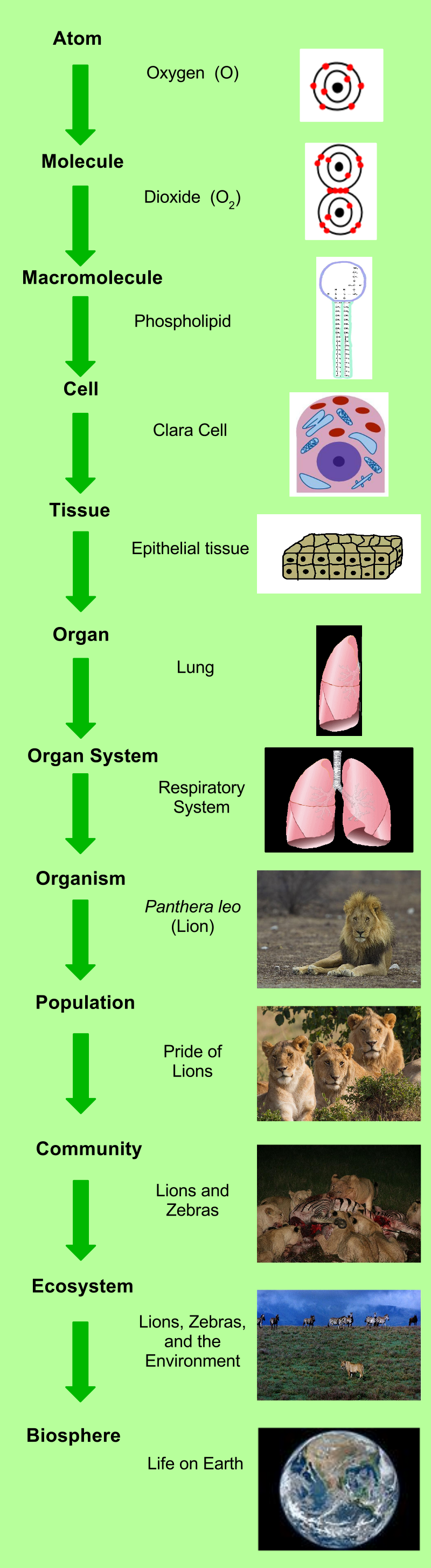
Living things are highly organized and structured, following a hierarchy of scale from small to large (Figure 1). The atom is the smallest and most fundamental unit of matter. It consists of a nucleus surrounded by electrons. Atoms combine to form molecules, which are chemical structures consisting of at least two atoms held together by a chemical bond. The chemical properties of molecules affect how they assemble into cells. All living things are made of one or more cells. In plants, animals, and other eukaryotic organisms, before the hierarchical level of cells, molecules come together in specific ways to create structures called organelles, which perform specific functions. Details of organelles and cells are discussed in more detail in the section 2.3.2.
In most multicellular organisms, cells combine to make tissues, which are groups of similar cells carrying out the same function. Organs are collections of tissues grouped together based on a common function. Organs are present not only in animals but also in plants. An organ system is a higher level of organization that consists of functionally related organs. For example vertebrate animals have many organ systems, such as the circulatory system that transports blood throughout the body and to and from the lungs; it includes organs such as the heart and blood vessels. Organisms are individual living entities. For example, each tree in a forest is an organism.
The levels of biology from organism to the biosphere are also called the ecological levels of organization. All the individuals of a species living within a specific area are collectively called a population. A community is the set of different populations inhabiting a common area. For instance, all of the trees, flowers, insects, and other populations in a forest form the forest’s community. The forest itself is an ecosystem. An ecosystem consists of all the living things in a particular area together with the abiotic, or non-living, parts of that environment such as nitrogen in the soil or rainwater. At the highest level of organization, the biosphere is the collection of all ecosystems, and it represents the zones of life on Earth. It includes land, water, and portions of the atmosphere. The biosphere is divided into biomes, which are discussed in more detail in sections 3.2 and 3.3.
Each level in the hierarchy represents an increase in organizational complexity, and is primarily composed of the previous level’s basic unit. E.g., a eukaryotic cell is primarily composed of organelles and molecules, the levels lower than that of “cell”. The basic principle behind the organization is the concept of emergence—the properties and functions found at a hierarchical level are not present and are even irrelevant at the lower levels.
2.3.2 Cell Theory
Close your eyes and picture a brick wall. What is the basic building block of that wall? It is a single brick, of course. Like a brick wall, your body is composed of basic building blocks and the building blocks of your body are cells. Your body has many kinds of cells, each specialized for a specific purpose. Just as a home is made from a variety of building materials, the human body is constructed from many cell types. For example, cells of the immune system fight invading bacteria, and red blood cells carry oxygen throughout the body. Each of these cell types plays a vital role during the growth, development, and day-to-day maintenance of the body. In spite of their enormous variety, however, all cells share certain fundamental characteristics.
The microscopes we use today are far more complex than those used in the 1600s by Antony van Leeuwenhoek, a Dutch shopkeeper who had great skill in crafting lenses. Despite the limitations of his now-ancient lenses, van Leeuwenhoek observed the movements of single-celled organism and sperm, which he collectively termed “animalcules.” In a 1665 publication called Micrographia, experimental scientist Robert Hooke coined the term “cell” (from the Latin cella, meaning “small room”) for the box-like structures he observed when viewing cork tissue through a lens. In the 1670s, van Leeuwenhoek discovered bacteria and protozoa. Later advances in lenses and microscope construction enabled other scientists to see different components inside cells.
By the late 1830s, botanist Matthias Schleiden and zoologist Theodor Schwann were studying tissues and proposed the unified cell theory, which states that all living things are composed of one or more cells, that the cell is the basic unit of life, and that all new cells arise from existing cells. These principles still stand today. There are many types of cells, and all are grouped into one of two broad categories: prokaryotic and eukaryotic. Animal, plant, fungal, and protist cells are classified as eukaryotic, whereas bacteria and archaea cells are classified as prokaryotic.
All cells share four common components: 1) a plasma membrane, an outer covering that separates the cell’s interior from its surrounding environment; 2) cytoplasm, consisting of a jelly-like region within the cell in which other cellular components are found; 3) DNA, the genetic material of the cell; and 4) ribosomes, particles that synthesize proteins. However, prokaryotes differ from eukaryotic cells in several ways.
Components of Prokaryotic Cells
A prokaryotic cell is a simple, single-celled (unicellular) organism that lacks a nucleus, or any other membrane-bound organelle. We will shortly come to see that this is significantly different in eukaryotes. Prokaryotic DNA is found in the central part of the cell: a darkened region called the nucleoid (Figure 2). Prokaryotes are divided into two domains of life: Bacteria and Archaea (see more in Section 5.1). Most of our current knowledge of prokaryotes is derived from studying bacteria. Note that the composition of the cell membrane and cell wall are different among the larger groupings of organisms (meaning archaea vs. bacteria vs. eukaryotic cells).
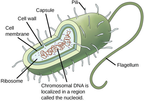
All bacteria have a cell wall in addition to their cell membrane with the specific compound called peptidoglycan (molecules comprised of sugars and amino acids) and many have a polysaccharide capsule outside the cell wall. The cell wall acts as an extra layer of protection, helps the cell maintain its shape, and prevents dehydration. The capsule typically enables the cell to attach to surfaces in its environment, and can be the specific feature that bothers the human immune system. Some prokaryotes have flagella, pili, or fimbriae. Flagella are used for locomotion. Pili are used to exchange genetic material during a type of reproduction called conjugation. Fimbriae are protein appendages used by bacteria to attach to other cells.
Eukaryotic Cells
A eukaryotic cell is a cell that has a membrane-bound nucleus and other membrane-bound compartments called organelles. Organelles are either separately enclosed within their own lipid bilayers (also called membrane-bound organelles) or are spatially distinct functional units without a surrounding lipid bilayer (non-membrane bound organelles). The word “organelle” means “little organ,” and, as already mentioned, organelles have specialized cellular functions, just as the organs of your body have specialized functions (see Figure 5 and 6). The word eukaryotic means “true kernel” or “true nucleus,” alluding to the presence of the membrane-bound nucleus in these cells.
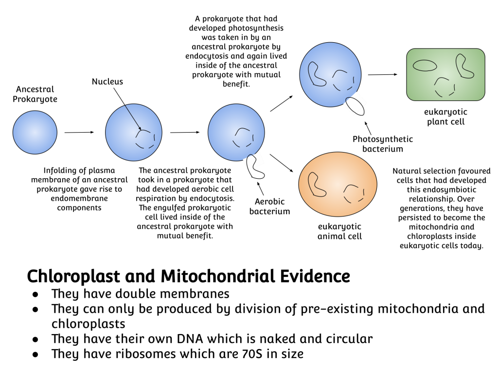
Two specific organelles in eukaryotes are thought to have originated through symbiogenesis: mitochondria and chloroplasts. The symbiogenesis theory proposes that these organelles evolved from certain types of bacteria that early eukaryotic cells engulfed through phagocytosis (Figure 3). These cells and the bacteria trapped inside them entered an endosymbiotic relationship, meaning that the bacteria took up residence and began living exclusively within the eukaryotic cells. Mitochondria and chloroplasts both have double membranes and separate DNA from than in the cell’s nucleus.
By Eosmanov – Own work, CC BY-SA 4.0.
Cell Size
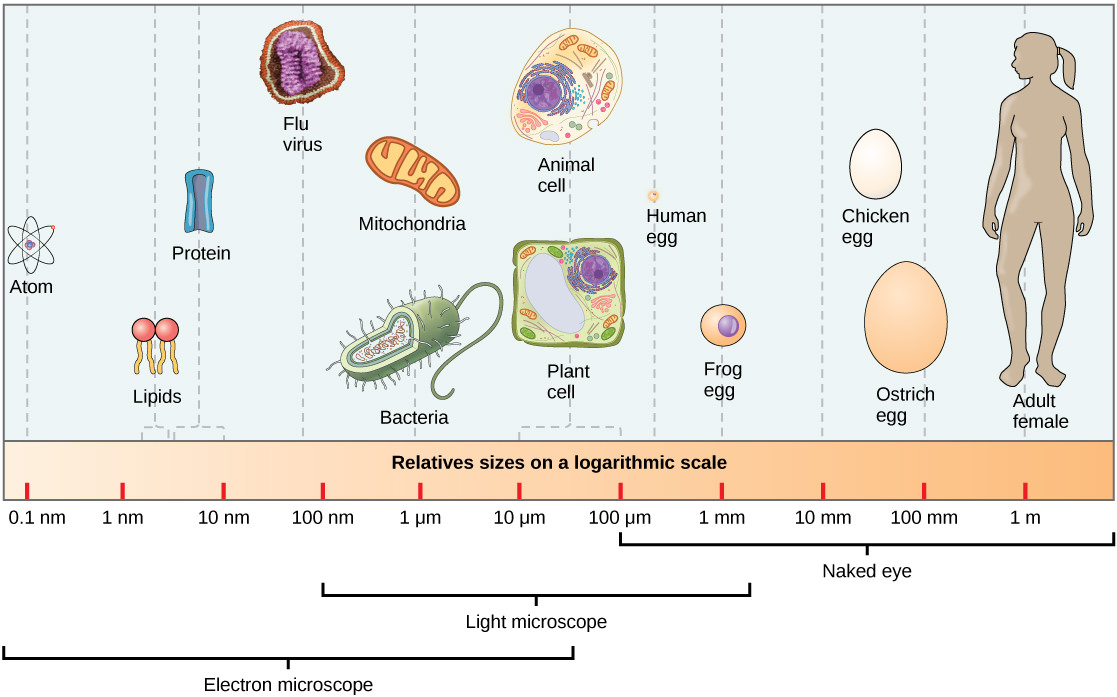
In general, cell size is limited because volume increases much more quickly than does cell surface area. As a cell becomes larger, it becomes more and more difficult for the cell to acquire sufficient materials to support the processes inside the cell, because the relative size of the surface area through which materials must be transported declines. At 0.1–5.0 µm in diameter, most prokaryotic cells are significantly smaller than eukaryotic cells, which have diameters ranging from 10–100 µm (Figure 4). The small size of prokaryotes allows ions and organic molecules that enter them to quickly spread to other parts of the cell. Similarly, any wastes produced within a prokaryotic cell can quickly move out. Larger eukaryotic cells have evolved different structural adaptations to enhance cellular transport with the most basic functions being to bring energy-providing molecules and to carry away waste products. Indeed, the large size of these cells would not be possible without these adaptations.
2.3.3 Chloroplasts and Mitochondria

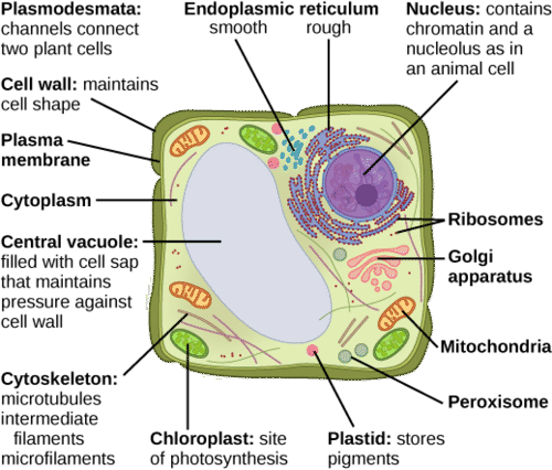
Despite their fundamental similarities, there are some striking differences between animal and plant cells (Figures 5 and 6). Animal cells have centrioles, centrosomes, and lysosomes, whereas plant cells do not. Plant cells have a rigid cell wall that is external to the plasma membrane, chloroplasts, plasmodesmata, and plastids used for storage, and a large central vacuole, whereas animal cells do not. These fundamental differences in cell membrane and cell wall connect to larger concepts like the digestibility of food.
Chloroplasts
One major difference between algae/plants and animals is that plants/algae are able to make their own food, like glucose, whereas animals must obtain food by consuming other organisms. Which leads us to this topic and subsection.
Chloroplasts are a particularly important type of organelle because they perform photosynthesis. Photosynthesis forms the foundation of food chains in most ecosystems. Chloroplasts are only found in eukaryotic cells such as plants and algae (some prokaryotic cells can also perform photosynthesis, but the majority of photosynthesis is performed by eukaryotic cells and organisms). During photosynthesis, carbon dioxide, water, and light energy are used to make glucose and molecular oxygen.
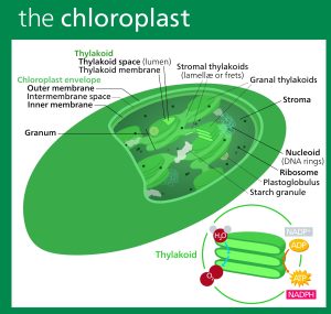
Chloroplasts have outer and inner membranes, but within the space enclosed by a chloroplast’s inner membrane is a set of interconnected and stacked, fluid-filled membrane sacs called thylakoids (Figure 7). Each stack of thylakoids is called a granum (plural = grana). The fluid enclosed by the inner membrane and surrounding the grana is called the stroma. The inner membranes help organize the reactions that are part of photosynthesis, and moving electrons across the inner membrane is a fundamental way that energy is transformed from solar energy into chemical energy.
A few common themes in biology are demonstrated in chloroplasts. First is that form and function are interrelated. Each structure within the chloroplast has an important function, which is enabled by its particular shape. For example, the membrane-rich stacks of the thylakoids provide ample surface area to embed the proteins and pigments that are vital to photosynthesis. Second is the importance of surface area in organization of organisms.
Mitochondria
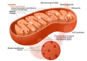
Most eukaryotic cells, both plants and animals, have mitochondria that function as a “powerhouse” to provide the cell with enough energy in the form of ATP to function. Mitochondria are important organelles thought to originate as separately living prokaryotic cells (like chloroplasts). Similar to chloroplasts, mitochondria have a high amount of membrane surface area regulating the interior of the mitochondria (Figure 8). These membranes control the movement of ions–most importantly, electrons and H+ cations–that allow the aerobic respiration reactions to break down complex carbon molecules like glucose into simple carbon dioxide molecules and transform the energy from one chemical bond to a different one (held in ATP).
In the next section, we will see these two chemical reactions in more detail: photosynthesis and aerobic respiration.
Attribution
Essentials of Environmental Science by Kamala Doršner is licensed under CC BY 4.0. Modified by Matthew R. Fisher and Joni Baumgarten.
Levels of Organization of Living Things by Open Stax is licensed under CC BY 4.0. Modified by Matthew R. Fisher and Joni Baumgarten.
Biological organisation by Wikipedia is licensed under CCA-SA 3.0. Modified from original by Joni Baumgarten.
Organelle by Wikipedia is licensed under CCA-SA 3.0. Modified from original by Joni Baumgarten.
