
The Digestive System
We just learned that our body is composed of billions of cells. To function, these cells need essential nutrients—carbohydrates, proteins, fats, vitamins, and minerals—which we obtain from foods. However, before our cells can access these nutrients, foods need to be broken down or digested into their simplest units, so that the nutrients can be absorbed and enter the bloodstream. Digestion is a complex process that involves many organs and chemicals, as we’ll explore on this page.
An Overview of the Organs Involved in Digestion
The function of the digestive system is to break down the foods you eat, release their nutrients, and absorb those nutrients into the body. Although the small intestine is the workhorse of the system where the majority of digestion and absorption occurs, each of the digestive system organs makes a vital contribution to this process.
The easiest way to understand the digestive system is to divide its organs into two main categories: the gastrointestinal tract (GI tract) and the accessory organs.
- The GI tract is a one-way tube about 25 feet in length, beginning at the mouth and ending at the anus. Between these two points, the GI tract also contains the pharynx, esophagus, stomach, small and large intestines, and the rectum. The small intestine is comprised of three parts: the duodenum, the jejunum, and the ileum. The large intestine, also called the colon, is similarly divided into three sections: the ascending colon, transverse colon, and descending colon. Both the mouth and anus are open to the external environment; thus, food and wastes within the GI tract are technically considered to be outside the body. Only through the process of absorption do the nutrients in food enter into and nourish the body’s “inner space.”
- Accessory organs, despite their name, are critical to the function of the digestive system. They are considered accessory organs since they are not actually part of the intestinal tract itself, but have ducts that deliver digestive juices into the tract to help aid in digestion. There are four accessory organs: the salivary glands, liver, gallbladder, and pancreas. All of these organs secrete fluids containing a variety of chemicals such as enzymes and acids that aid in digestion.
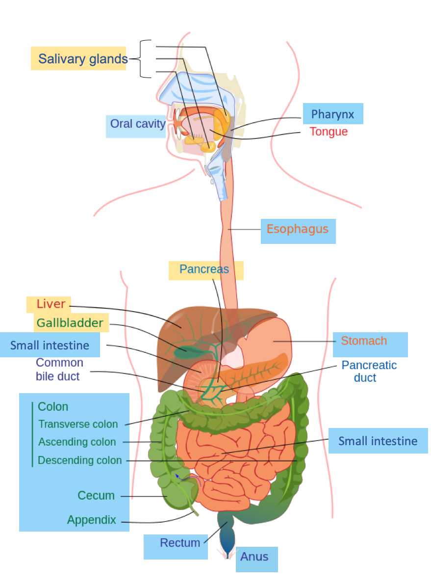
Figure 3.9. An overview of the organs involved in digestion. The parts of the GI tract are highlighted in blue, and the accessory organs are highlighted in yellow.
An Overview of the Digestive Process
The process of digestion includes five main activities: ingestion, mechanical digestion, chemical digestion, absorption, and excretion.
The first of these processes, ingestion, refers to the entry of food into the GI tract through the mouth. There, the food is chewed and mixed with saliva, which contains enzymes that begin breaking down the carbohydrates and lipids in food. Mastication (chewing) increases the surface area of the food and allows for food to be broken into small enough pieces to be swallowed safely.
Food (now called a bolus since it has been chewed and moistened) leaves the mouth when the tongue and pharyngeal muscles propel the bolus into the esophagus. The bolus will travel down the esophagus through an involuntary process called peristalsis. Peristalsis consists of sequential, alternating waves of contraction and relaxation of the smooth muscles in the GI tract, which act to propel food along (Figure 3.10). These waves also play a role in mixing food with digestive juices. Peristalsis is so powerful that foods and liquids you swallow enter your stomach even if you are standing on your head.
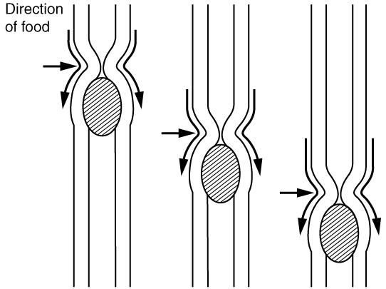
Figure 3.10. Peristalsis moves food through the digestive tract with alternating waves of muscle contraction and relaxation.
Digestion includes both mechanical and chemical processes. Mechanical digestion is a purely physical process of making food particles smaller to increase both surface area and mobility. Mechanical digestion does not change the chemical nature of the food. It includes mastication, tongue movements that help break food into smaller bits and mix it with saliva, mixing and churning of the stomach to further break food apart and expose more of its surface area to digestive juices, and peristalsis to help move food along the intestinal tract. Segmentation is also an example of mechanical digestion. Segmentation, which occurs mainly in the small intestine, consists of localized contractions of circular muscle of the GI tract. These contractions isolate small sections of the intestine, moving their contents back and forth while continuously subdividing, breaking up, and mixing the contents. By moving food back and forth in the intestinal tract, segmentation mixes food with digestive juices and facilitates absorption.
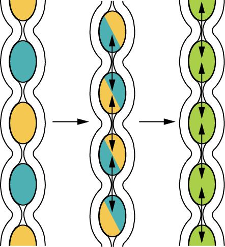
Figure 3.11. Segmentation separates chyme and then pushes it back together, mixing it and providing time for digestion and absorption.
In chemical digestion, digestive secretions that contain enzymes start to break down the macronutrients into their chemical building blocks (for example, starch into glucose). Enzymes are chemicals that help speed up or facilitate chemical reactions in the body. They bring together two compounds to react, without undergoing any changes themselves. For example, the main chemical reaction in digestion is hydrolysis.Hydrolysis is the splitting of one molecule into two with the addition of water. For example, the sugar sucrose (a double sugar) needs to be broken down to its building blocks, glucose and fructose (both single sugars), before it can be absorbed. This breakdown happens through hydrolysis, and the enzyme, sucrase, brings together the sucrose molecule and the water molecule to react. This process is illustrated in the following animation.
Video: Enzyme Action and the Hydrolysis of Sucrose by McGraw-Hill Animations, YouTube (June 3, 2017). 1:46 minutes.
Nutrients are of little to no value to the body unless they enter the bloodstream. This occurs through the process of absorption, which takes place primarily within the small intestine. There, most nutrients are absorbed from the lumen (or inside space) of the GI tract into the bloodstream. Larger lipids are absorbed into lymph but eventually enter the bloodstream as well.
In excretion, the final step of digestion, undigested materials are removed from the body as feces. The feces is stored in the rectum until it leaves the body through the anus.
Functions of the Digestive Organs
Now that you have an overview of the digestive organs and the digestive process, let’s discuss in more detail what types of mechanical and chemical digestion take place in each of the organs of the GI tract. Let’s imagine eating a peanut butter and jelly sandwich that contains carbohydrates, proteins, fats, vitamins, and minerals. How does each organ participate in breaking this sandwich down into units that can be absorbed and utilized by cells throughout the body?
Mouth
Ingestion of the peanut butter and jelly sandwich happens in the mouth or oral cavity. This is where mechanical and chemical digestion also begin. Teeth physically crush and grind the sandwich into smaller particles and mix the food particles with saliva. Salivary amylase (a digestive enzyme) is secreted by salivary glands (salivary glands produce saliva which is a mixture of water, enzymes, and other chemicals) and begins the chemical breakdown of carbohydrates in the bread, while lingual lipase (another digestive enzyme) starts the chemical breakdown of triglycerides (the main form of fat in food) in the peanut butter.
Esophagus
The esophagus is a muscular tube that transports food from the mouth to the stomach. No chemical digestion occurs while the bolus is mechanically propelled through this tube by peristalsis.
Stomach
The stomach is an expansion of the GI tract that links the esophagus to the first part of the small intestine (the duodenum). The empty stomach is only about the size of your fist but can stretch to hold as much as 4 liters of food and fluid—more than 75 times its empty volume—and then return to its resting size when empty. An important function of the stomach is to serve as a temporary holding chamber. You can ingest a meal far more quickly than it can be digested and absorbed by the small intestine. Thus, the stomach holds food and secretes only small amounts into the small intestine at a time. (The length of time food spends in the stomach varies by the macronutrient composition of the meal. A high-fat or high-protein meal takes longer to break down than one rich in carbohydrates. It usually takes a few hours after a meal to empty the stomach contents completely into the small intestine.)
When the peanut butter and jelly sandwich enters the stomach, a highly muscular organ, powerful peristaltic contractions help mash, pulverize, and churn it into chyme. Chyme is a semiliquid mass of partially digested food along with gastric juices secreted by cells in the stomach. These gastric juices contain hydrochloric acid, which lowers the pH of the chyme in the stomach. This acidic environment kills many bacteria or other germs that may have been present in the food, and it causes the three-dimensional structure of dietary proteins to unfold. Gastric juices also contain the enzyme pepsin, which begins the chemical breakdown of proteins in the peanut butter and bread. Gastric lipase continues the breakdown of fat from the peanut butter.
Small Intestine
Chyme released from the stomach enters the small intestine, where most digestion and absorption occurs. The small intestine is divided into three parts, all part of one continuous tube: the duodenum, the jejunum, and the ileum.
Once the chyme enters the duodenum (the first segment of the small intestine), the pancreas and gallbladder are stimulated to release juices that aid in digestion. The pancreas (located behind the stomach) produces and secretes pancreatic juices which consist mostly of water, but also contain bicarbonate that neutralizes the acidity of the stomach-derived chyme and enzymes that further break down proteins, carbohydrates, and lipids. The small intestine’s absorptive cells also synthesize digestive enzymes that aid in the breakdown of sugars and proteins.
The gallbladder (a small sac located behind the liver) stores, concentrates, and secretes a fluid called bile that helps to digest fats. Bile is made in the liver and stored in the gallbladder. Bile is an emulsifier; it acts similar to a detergent (that would remove grease from a frying pan) by breaking large fat droplets into smaller fat droplets so they can mix with the watery digestive juices.
Peristalsis and segmentation control the movement and mixing of chyme through the small intestine. As in the esophagus and stomach, peristalsis consists of circular waves of smooth muscle contractions that propel food forward. Segmentation helps to mix food with digestive juices and facilitates absorption.
Nutrient absorption takes place mainly in the latter part of the small intestine, the ileum. The small intestine is perfectly structured for maximizing nutrient absorption. Its surface area is greater than 200 square meters—about the size of a tennis court! The large surface area is due to the multiple levels of folding, villi, and microvilli that cover the internal tissue of the small intestine. Villi are tiny finger-like projections that are covered with enterocytes or absorptive cells. The absorptive cell membrane is made of even smaller projections, called microvilli (Figure 3.12). These microvilli are referred to collectively as the brush border since their appearance resembles the bristles on a brush.
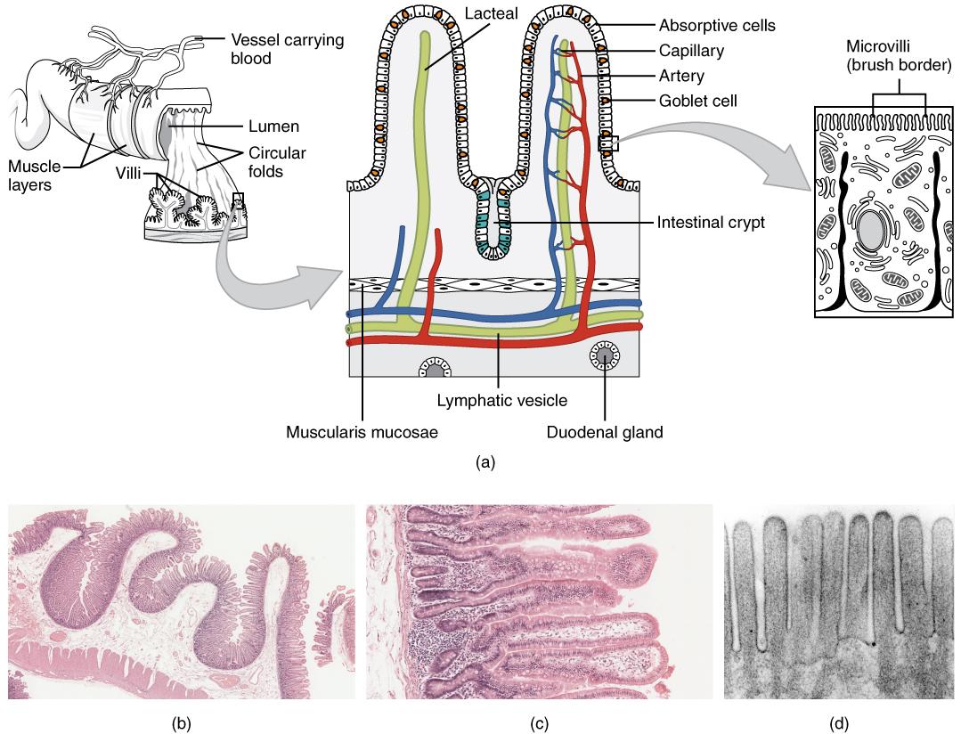
Figure 3.12. Histology of the small intestine. (a) The absorptive surface of the small intestine is vastly enlarged by the presence of circular folds, villi, and microvilli. (b) Micrograph of the circular folds. (c) Micrograph of the villi. (d) Electron micrograph of the microvilli.
Digested nutrients are absorbed into either capillaries or lymphatic vessels contained within each villus. Amino acids (from protein digestion), small fatty acids (from triglyceride digestion), sugars (from carbohydrate digestion), water-soluble vitamins, and minerals are transported from the intestinal cells into the bloodstream through capillaries. The larger fatty acids, fat-soluble vitamins, and other lipids (that are packaged in lipid transport particles) are transported first through lymphatic vessels and then eventually meet up with the blood. Water-soluble nutrients that enter the bloodstream are transported directly to the liver where the liver processes, stores, or releases these nutrients to other body cells.
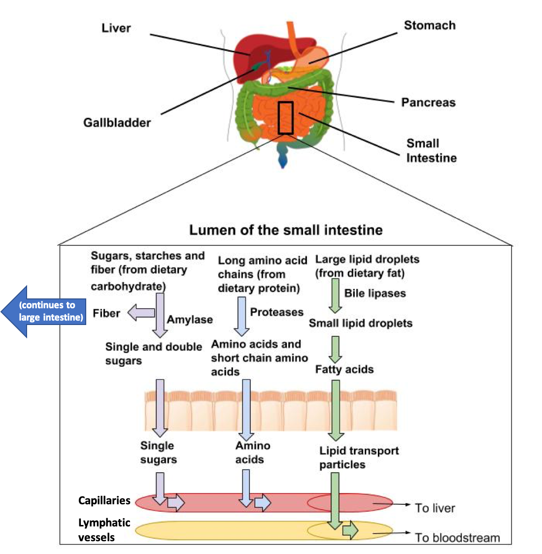
Figure 3.13. The digestion and absorption of nutrients in the small intestine.
Large Intestine
Most of the nutrients from the peanut butter and jelly sandwich have now been digested and absorbed. Any components that still remain (usually less than ten percent of food consumed) and the indigestible fiber move from the small intestine to the large intestine (colon). A main task of the large intestine is to absorb much of the remaining water. Water is present not only from the solid foods and beverages consumed, but also the digestive juices released by the stomach and pancreas. As water is reabsorbed, liquid chyme becomes a semisolid, referred to as feces. Feces is composed of undigested food residues, unabsorbed digested substances, millions of bacteria, old cells from the lining of the GI tract, inorganic salts, and enough water to let it pass smoothly out of the body.
Feces is stored in the rectum (a temporary holding area) until it is expelled through the anus via defecation. No further chemical breakdown of food takes place in the large intestine except that accomplished by the bacteria that inhabit this portion of the GI tract. There are trillions of bacteria residing in the large intestine (referred to as the bacterial flora), exceeding the total number of cells in the human body. This may seem rather unpleasant, but the great majority of bacteria in the large intestine are harmless and many are even beneficial—facilitating chemical digestion and absorption, improving immune function, and synthesizing vitamins such as biotin, pantothenic acid, and vitamin K.
The figure below summarizes the functions of the digestive organs.
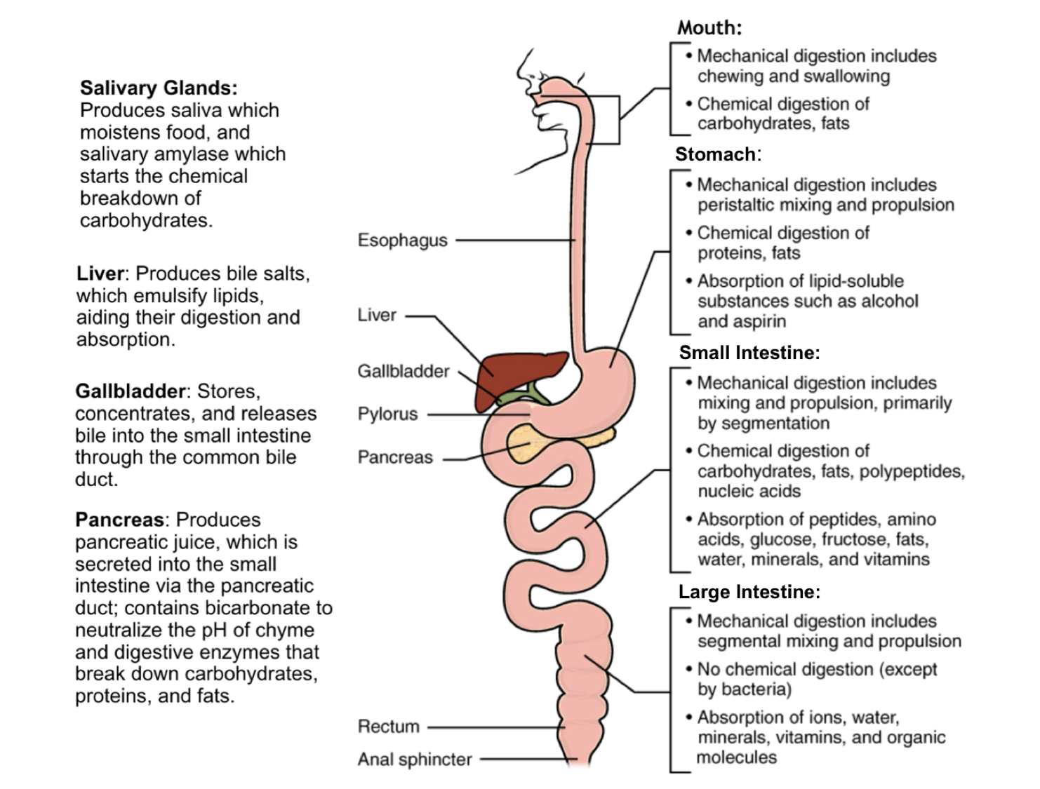
Figure 3.14. Summary of digestion and absorption. Digestion begins in the mouth and continues as food travels through the small intestine. Most absorption occurs in the small intestine.
Video: “The Digestive System” by National Geographic, YouTube (November 26, 2012), 5:07 minutes.
Self-Check:
Attributions:
- “The Digestive System,” unit 23 from J. Gordon Betts, Kelly A. Young, James A. Wise, Eddie Johnson, Brandon Poe, Dean H. Kruse, Oksana Korol, Jody E. Johnson, Mark Womble, Peter DeSaix, Anatomy and Physiology, CC BY 4.0
- University of Hawai‘i at Mānoa Food Science and Human Nutrition Program, “The Digestive System,” CC BY-NC 4.0
Images:
- Figure 3.9. “GI tract and accessory organs” by Alice Callahan is licensed under CC BY 4.0; edited from “Digestive system diagram edit” by Mariana Ruiz, edited by Joaquim Alves Gaspar, Jmarchn is in the Public Domain
- Figure 3.10. “Peristalsis” by J. Gordon Betts, Kelly A. Young, James A. Wise, Eddie Johnson, Brandon Poe, Dean H. Kruse, Oksana Korol, Jody E. Johnson, Mark Womble, Peter DeSaix, Anatomy and Physiology, OpenStax, licensed under CC BY 4.0
- Figure 3.11. “Segmentation” by OpenStax College is licensed under CC BY 3.0
- Figure 3.12. “Histology Small Intestines” by OpenStax College is licensed under CC BY 3.0
- Figure 3.13. “Absorption of Nutrients” by Tamberly Powell is licensed under CC BY 4.0; edited from University of Hawai‘i at Mānoa Food Science and Human Nutrition Program, “The Digestive System,” CC BY-NC 4.0
- Figure 3.14. “Functions of the Digestive Organs” by Tamberly Powell is licensed under CC BY 4.0; edited from “Figure 23.28 Digestion and Absorption” by J. Gordon Betts, Kelly A. Young, James A. Wise, Eddie Johnson, Brandon Poe, Dean H. Kruse, Oksana Korol, Jody E. Johnson, Mark Womble, Peter DeSaix , Anatomy and Physiology, OpenStax, licensed under CC BY 4.0
A one-way tube about 25 feet in length, beginning at the mouth and ending at the anus; where digestion takes place.
Organs that are not part of the intestinal tract itself, but have ducts that deliver digestive juices into the tract to help aid in digestion; includes the salivary glands, liver, gallbladder, and pancreas.
The first process of digestion; the entry of food into the GI tract through the mouth.
Chewing; increases the surface area of the food and allows for food to be broken into small enough pieces to be swallowed safely.
What food is referred to once it has been chewed and moistened.
Sequential, alternating waves of contraction and relaxation of the smooth muscles in the GI tract; acts to propel food along the GI tract.
The physical process of making food particles smaller to increase both surface area and mobility without changing the chemical nature of the food (e.g., mastication and peristalsis).
Consists of localized contractions of circular muscle of the GI tract; isolates small sections of the intestine, moving contents back and forth while continuously subdividing, breaking up, and mixing the contents; mixes food with digestive juices and facilitates absorption.
The breakdown of macronutrients into their chemical building blocks (e.g., starch into glucose) with the aid of enzymes.
Proteins that help speed up or facilitate chemical reactions in the body; they bring together two compounds to react, without undergoing any changes themselves.
The splitting of one molecule into two with the addition of water; the main chemical reaction in digestion.
The process of nutrients traveling from the lumen of the GI tract to the blood or lymph after digestion.
The interior space of the GI tract where digestion and absorption occur.
The final step of digestion; undigested materials are removed from the body.
Undigested materials that are stored in the anus until they are excreted from the body.
The oral cavity where ingestion and mastication occur.
A digestive enzyme produced by the salivary glands; starts the chemical breakdown of starch or amylose.
The glands that make and secrete saliva.
A mixture of water, enzymes, and other chemicals secreted from the salivary glands into the mouth.
An enzyme produced by cells on the tongue; begins the chemical breakdown of triglycerides.
A muscular tube that transports food from the mouth to the stomach.
An expansion of the GI tract that links the esophagus to the first part of the small intestine; aids in both mechanical and chemical breakdown of food.
A semiliquid mass of partially digested food and gastric juices.
An acid that is a component of gastric juices; creates an acidic environment in the stomach, killing bacteria and aiding in protein digestion.
An enzyme found in gastric juices; aids in the chemical breakdown of proteins.
An enzyme produced by cells of the stomach; aids in the chemical breakdown of triglycerides.
A part of the GI tract that lies between the stomach and the large intestine; where most digestion and nutrient absorption occurs.
The first segment of the small intestine.
An accessory organ located behind the stomach; produces and secretes pancreatic juices, which contain bicarbonate that neutralizes the acidity of the stomach-derived chyme and enzymes that further break down proteins, carbohydrates, and lipids.
An accessory organ located behind the liver; stores, concentrates, and secretes bile.
An accessory organ located just under the rib cage on the right side of the abdomen; produces bile and helps get rid of toxins.
A chemical made by the liver and stored in the gallbladder; acts as an emulsifier, which allows fat droplets to mix with the watery digestive juices in the small intestine.
The final segment of the small intestine, where the majority of absorption occurs.
Tiny, finger-like projections that cover the lining of the small intestine to increase surface area.
Absorptive cells that line the small intestine.
Hair-like projections that line the enterocytes’ cell membrane to increase surface area.
Another name for microvilli, because their appearance resembles the bristles on a brush.
The part of the GI tract that lies between the small intestine and the anus; water absorption occurs here.
The last part of the GI tract; serves as a temporary holding area for feces.
