9 Colony Morphology
Emilie Miller, Ph.D
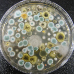
On agar plates, bacteria grow in collections of cells called colonies. Each colony arises from a single bacterium or a few bacteria (CFU). Although individual cells are too small to be viewed with the unaided eye, masses of cells can be observed. Colonies can have different forms, margins, elevations and colors. Observing colony characteristics is one piece of information that microbiologists can use to identify unknown bacteria. (Petersen, 2016)
In this exercise you will be drawing colonies and describing each type’s size, shape, elevation, margin, color and texture. Keep in mind that the colony characteristics of a microbe may change depending on the medium, time and temperature of incubation. The medium supplies the nutrients and other materials for the cell to use. Along with the size of individual cells, the colony size depends on the speed at which the cells divide. This is determined by the organism’s inherent cell cycle, the availability of nutrients and the organism’s optimal growth temperature.

Size can be actually measured in millimeters or described as “pin point.”
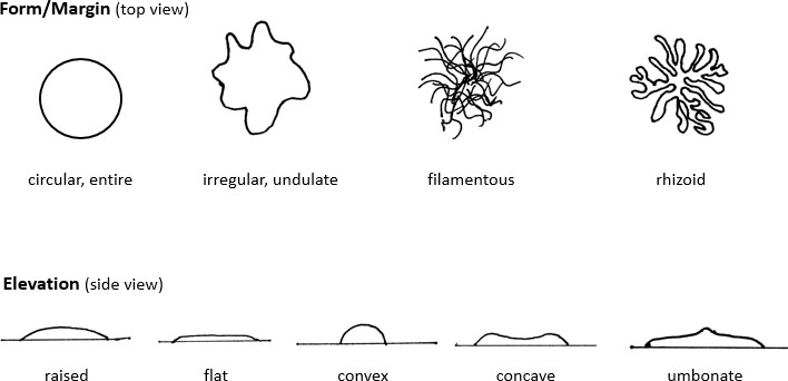
Colony form means the shape and can be circular, irregular, or rhizoid (branched). This is the cumulative (macroscopic) effect of the microscopic cellular shape and arrangement.
Elevation refers to the cross sectional view or profile of the colony. It can be raised, flat, convex, concave or umbonate.
Margin describes the edge. A smooth edge is called entire. Other margins are undulate (an irregular, wavy edge), lobed (more pronounced wavy edge) or spreading (no distinct colonies).
Filamentous and rhizoid may also be used to describe the margin.
Colony color can be the result of the color of the actual cell, the result of pigments produced by the cell under certain conditions, or the interaction of certain cellular metabolites with components in the medium.
Texture can be dry, ridged or wavy, mucoid/shiny, dull, filamentous (hairy looking).
Procedure
- Observe one colony from each of the different pure cultures.
- Note the full organism name written in full scientific form.
- A sketch of the top view and the side view. For the side view, draw a straight line to represent the agar surface as in figure 1 above.
- Use relevant terms to describe the size (measure, if .5 mm or larger), color, form, elevation and texture of each.
References:
Petersen, J. a. (2016). Laboratory Exercises in Microbiology: Discovering the Unseen World Through Hands-On Investigation. CUNY Academic Works. Retrieved from http://academicworks.cuny.edu/qb_oers/16

