6 Microscopy Assignment
Emilie Miller, Ph.D
The human mouth is home to numerous microbes, which persist no matter how many times you brush your teeth and use mouthwash. Since these microbes generally inhabit the surface layers of the oral mucosa, we humans have evolved ways to keep their numbers under control, including producing antibacterial chemicals in saliva and constantly turning over the outer layer of epithelial cells that line the inside of the mouth.
These are squamous epithelial cells that form the outermost layer of the oral mucosa. At high power, you should start to see small cells on the surface of the larger epithelial cells. With the oil immersion objective lens, you will be able to tell the smaller cells are bacteria.
- Gently swab your inner cheek with a cotton swab and swirl in a small spot on a clean slide. Flame your slide quickly over a bunsen burner to fix the cells. Apply a few drops of Crystal Violet to the cells then rinse the slide with water. Blot the slide dry with Bibulous Paper and then view your slides with your microscope working your way up through the objectives.
Draw and label your cheek cells and include the cell membrane, nucleus using the 40x objective: Total magnification: ______
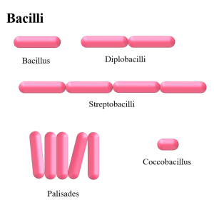
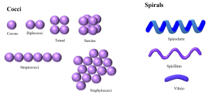
2. Use the 100x objective with oil to view bacteria that may have been fixed outside and inside your cheek cells: Draw and ID the shapes present using the terms: Cocci, Bacilli, Strepto, Staphlo. Get confirmation of your image from the instructor when you have it in view_______. Total magnification:______.