
Pupil Assessment
They say they eyes are the window to the soul; however, it would be more accurate to say they are a window to the brain. In a healthy eye and brain, the pupil responds to allow light waves to properly stimulate the nerves of the retina. The stimulation of nerves, called “rods” and “cones” within the retina is translated to signals along the optic nerve to the occipital lobe of the brain. Within low-light settings the pupil dilates, or “grows larger,” to let needed light stimulate the retina. In high-light settings the pupils constrict, become smaller, to limit the light and avoid damage to the sensitive nerves of the retina.
The pupil assessment may reveal the presence of certain drugs, or trauma to the eye or brain. Medications such as opiates can cause the pupils to constrict and be “pinpoint” and not reactive to light. Other drugs such as psychotropics, stimulants and alcohol can cause pupils to become dilated. A non-reactive, “fixed” or asymmetric, “blown” pupil is a late sign of increased intracranial pressure and may increase the EMT’s index of suspicion for trauma to the brain or eye, or may point to the presence of a stroke. Note that genetic blindness in eyes will, in most cases make the pupils unresponsive to light changes.
You may hear several similar mnemonics for what to assess when looking at the eyes. The authors recommend the use of a standardized pneumonic such as, “PERRL.” Whichever acronym you choose to work with, stick with it for your whole career.
P – Pupils
E – Equal
R – Round
R – Reactive
L – to Light
To assess pupil responsiveness:
-
- First look at the patient pupil without additional light to determine size of pupil.
- Use a pen light to shine light from the far corner of the eye to the bridge of the nose.
- Note – do not use a super-bright flashlight as the additional light can be painful for your patient.
- As you shine the light, look for changes in pupil size in both eyes. Eye response is connected and both pupils should respond to light in one eye.
- Pupil reactive rate of change is also noted.
- Perform this assessment in each eye.
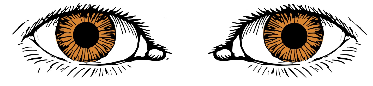
Fig 1. Normal pupils.
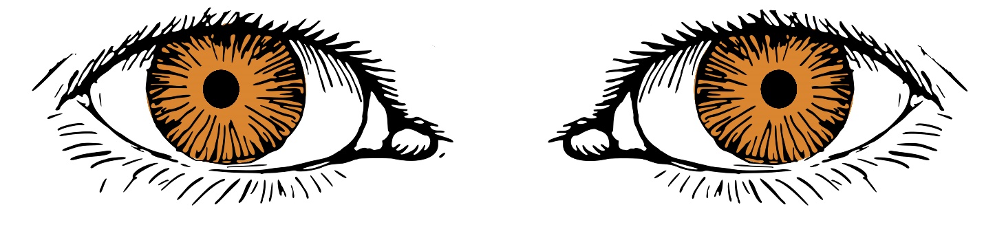
Fig 2. Pinpoint pupils
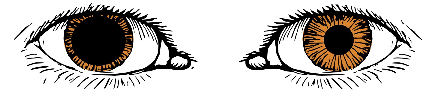
Fig 3. Blown pupils
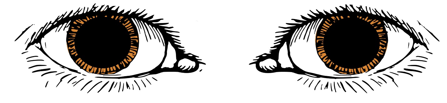
Fig 4. Dilated pupils
Images of pupils by Michaela Willi Hooper for Open Oregon, CC BY-NC-SA 4.0.
|
Pupil Assessment |
1 |
2 |
3 (Instructor) |
|
Initials |
|
|
|
The original copy of this book resides at openoregon.pressbooks.pub/emslabmanual. If you are reading this work at an alternate web address, it may contain content that has not been vetted by the original authors and physician reviewers.
