84 Comparing Meiosis and Mitosis

Video Transcript

In biology, there are often vocabulary terms that sound pretty similar.
Chromosome.
Chromatid.
Chromatin.
Transcription.
Translation.
Mitosis.
Meiosis.
You probably have encountered this.
When I was first learning about mitosis and meiosis, I learned them both separately first. And then I tried to figure out what was the same about them, what was different, why did they both matter? I would try to compare the stages by flipping through images.
You know what would have helped me? A side by side comparison. And that’s what this video is.
We assume you already have a background of mitosis and meiosis – if not, take a look at our videos on them – but this video is a side by side comparison. Presented in a split screen. Mitosis on the left. Meiosis on the right.
Both of these processes, along with the cytokinesis that follows them to split the cytoplasm, are involved in making new cells. Mitosis results in body cells. Meiosis results in sperm and egg cells, otherwise known as the fancy term, gametes.
Before we start mitosis and meiosis, let’s look at what you start with. Your starting cell in both mitosis and meiosis is diploid, written here as 2n. That means it has 2 sets of chromosomes – in humans, that’s including one set of 23 chromosomes from mom and one set of 23 chromosomes from dad. 46 chromosomes total in humans.
During interphase, the cell duplicates the chromosomes. When you duplicate 46 chromosomes, you still say there are 46 chromosomes as the newly duplicated portion is still attached at the centromere region – but there are actually 92 chromatids. Interphase isn’t part of mitosis or meiosis, but it’s a really important phase because it duplicates chromosomes before we get started.
Just to point out, it’s really hard to draw 46 chromosomes which is how many humans have. We’re going to use 6 chromosomes in our diagrams when we illustrate what’s happening because it’s much easier to draw and visualize. Oh and just a fun fact: some insects have 6 chromosomes. Like mosquitoes. Unfortunately, I am not a fan of mosquitoes. But mosquitoes do mitosis and meiosis too.
When learning the stages, we give the acronym PMAT which is helpful for understanding the stages. Both mitosis and meiosis go through these stages, but meiosis goes through them twice and therefore has a number next to each PMAT stage. We’re going to show some basic events for each PMAT stage, but please know there is way more detail to explore than what we can include in this quick video.
Prophase in mitosis. Remember that “pro” can mean “before” and this stage comes before the others. The chromosomes are visible; in fact, we say they’re condensing which means they are thickening.
Prophase I in meiosis. Happening here too, but the chromosomes are actually going to match up with their homologous pairs. The word homologous means that the chromosomes are approximately the same size and that they contain the same types of genes in the same locations. With each pair, one came from mom and one came from dad. In this formation, chromosomes can transfer their genetic information and exchange it between each other. It’s called crossing over! It can make for what we call recombinant chromosomes.
Metaphase in mitosis. The nuclear envelope which had surrounded the nucleus was already disassembled before metaphase started. For metaphase, I like to remember the M for middle because in this stage the chromosomes line up in the middle of the cell in a single file line.
Metaphase I in meiosis. The chromosomes are in the middle as well, but they’re still going to be in pairs in the middle of the cell so it’s not a single file line.
Anaphase in mitosis. I like to think as the A is for “away.” The chromatids are pulled away by the work of the spindles. They are moving to the opposite sides of the cell.
Anaphase I in meiosis. Same thing but in this case, it’s the chromosomes- not chromatids- being pulled away to opposite sides of the cell.
Telophase in mitosis and telophase I in meiosis. The chromosomes are at the complete opposite ends and new nuclei are forming on each side to make these two new cells. And they are starting to surround the chromosomes on both sides as this will eventually form 2 cells.
Cytokinesis follows to split the cytoplasm to complete the actual dividing of the cell.
So at the end of mitosis and cytokinesis, we end with two identical, diploid cells. In humans, they would both have 46 chromosomes. This is great for organism growth – growing requires making more cells after all – or replacing damaged cells.
On to meiosis II!
Prophase II. Chromosomes condensing in both cells. It’s not going to be as eventful as it was in prophase I because they are not going to have homologous pairs and crossing over.
Metaphase II. M for middle, but this time, the chromosomes are in a single file line. Similar to how metaphase looked in mitosis.
Anaphase II. Think A for away. This time, though, it’s actually the chromatids that are getting pulled away.
Telophase II. Chromosomes are at the complete opposite ends and new nuclei are forming on each side to make new cells.
Cytokinesis will follow meiosis II to completely split the cytoplasm.
We are now finished with meiosis: and we end with four non-identical cells. Gametes. Males make sperm cells in meiosis and females make egg cells in meiosis. These gametes are haploid, meaning they have half the number of chromosomes as the original starting cell. In the case of humans, the resulting cells would each have 23 chromosomes. By the way, when a sperm and egg cell combine, it results in a diploid cell, a fertilized egg otherwise known as a zygote, which will then start a series of divisions using mitosis to give rise to a brand new organism.
Well, that’s it for the Amoeba Sisters, and we remind you to stay curious!

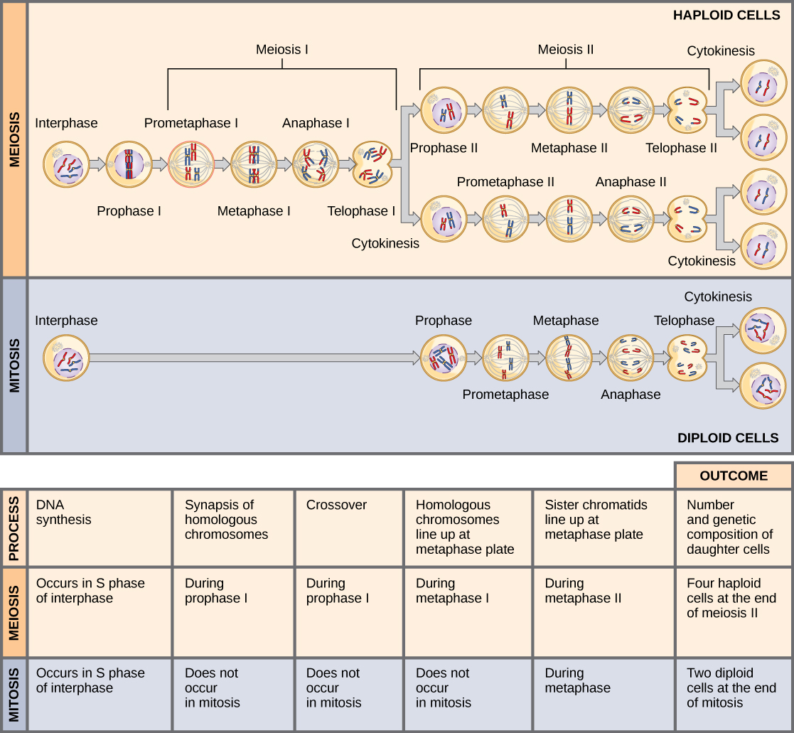
Mitosis and meiosis, which are both forms of division of the nucleus in eukaryotic cells, share some similarities, but also exhibit distinct differences that lead to their very different outcomes. Mitosis is a single nuclear division that results in two nuclei, usually partitioned into two new cells. The nuclei resulting from a mitotic division are genetically identical to the original. They have the same number of sets of chromosomes: one in the case of haploid cells, and two in the case of diploid cells. On the other hand, meiosis is two nuclear divisions that result in four nuclei, usually partitioned into four new cells. The nuclei resulting from meiosis are never genetically identical, and they contain one chromosome set only—this is half the number of the original cell, which was diploid.

The differences in the outcomes of meiosis and mitosis occur because of differences in the behavior of the chromosomes during each process. Most of these differences in the processes occur in meiosis I, which is a very different nuclear division than mitosis. In meiosis I, the homologous chromosome pairs become associated with each other, are bound together, experience crossover between homologous chromosomes, and line up in the center of the cell with spindle fibers from opposite spindle poles attached to each centromere. All of these events occur only in meiosis I, never in mitosis.

Homologous chromosomes move to opposite poles during meiosis I so the number of sets of chromosomes in each nucleus-to-be is reduced from two to one. For this reason, meiosis I is referred to as a reduction division. There is no such reduction in mitosis.

Meiosis II is much more similar to a mitotic division. In this case, duplicated chromosomes line up at the center of the cell. One sister chromatid is pulled to one pole and the other sister chromatid is pulled to the other pole. If it were not for the fact that there had been crossovers, the two products of each meiosis II division would be identical as in mitosis; instead, they are different because there has always been at least one crossover per chromosome. Meiosis II is not a reduction division because, although there are fewer copies of the genome in the resulting cells, there is still one set of chromosomes, as there was at the end of meiosis I.

Cells produced by mitosis will function in different parts of the body as a part of growth or replacing dead or damaged cells. Mitosis typically occurs in somatic cells, but they may be involved in asexual reproduction in some organisms. Cells produced by meiosis will only participate in sexual reproduction.

Meiosis and mitosis
Figure 84.1. Meiosis and mitosis are both preceded by one round of DNA replication; however, meiosis includes two nuclear divisions. The four daughter cells resulting from meiosis are haploid and genetically distinct. The daughter cells resulting from mitosis are diploid and identical to the parent cell.
Figure 84.1 Image Description

The processes of Meiosis and Mitosis are illustrated at the top of the diagram. Meiosis goes through two rounds of division. Meiosis I separates the homologous pairs of chromosomes and Meiosis II separates the sister chromatids. At the end of meiosis II, cytokinesis results in 4 haploid cells, each containing one copy of each chromosome. During Mitosis, only one division occurs. The sister chromatids are separated from each other. At the end of mitosis, cytokinesis results in 2 diploid cells, each containing two copies of each chromosome.

Process DNA Synthesis Synapsis of homolgous chromosomes Crossover Homologous Chromosomes line up at metaphase plate Sister chromatids line up at metaphase plate Number and genetic composition of daughter cells
Meiosis Occurs in S phase of interphase During prophase I During prophase I During metaphase I During metaphase II Four haploid cells at the end of meiosis II
Mitosis Occurs in S phase of interphase Does not occur Does not occur Does not occur During metaphase Two diploid cells at the end of mitosis

References

Unless otherwise noted, images on this page are licensed under CC-BY 4.0 by OpenStax.

OpenStax, Biology. OpenStax CNX. May 27, 2016. http://cnx.org/contents/s8Hh0oOc@9.10:1Q8z96mT@4/Meiosis

License

Icon for the Creative Commons Attribution 4.0 International License

MHCC Biology 112: Biology for Health Professions Copyright © 2019 by Lisa Bartee is licensed under a Creative Commons Attribution 4.0 International License, except where otherwise noted.

Share This Book