"

18 Comparing Prokaryotic and Eukaryotic Cells

Cells fall into one of two broad categories: prokaryotic and eukaryotic. Bacteria are classified as prokaryotes (pro– = before; –karyon– = nucleus). Animal cells, plant cells, fungi, and protists are eukaryotes (eu– = true).

Video Transcript

On Earth, there are two major types of cells: prokaryotic and eukaryotic.
Prokaryotic cells are the simplest and most ancient types of cells. They were the only form of life on Earth for billions of years before eventually giving rise to eukaryotic cells. If you take a look at prokaryotic and eukaryotic cells side by side, you can see a number of differences. To begin with, prokaryotic cells are a lot smaller than eukaryotic cells and have a simpler structure. But bear in mind that this simple structure is actually a good thing for prokaryotes, because it allows them to reproduce very quickly and very effectively.
If you were to look inside a prokaryotic cell, you would probably be surprised at how simple it is. For example, prokaryotic cells lack a nucleus. In fact, the name “prokaryote” actually gives you a hint about its structure. “Kary” means “kernel” or “nucleus” and “pro” means before. So basically, the word “prokaryote” means “before nucleus.”
The inside of the prokaryotic cell is basically an open unit with no compartments. There are no membrane-bound organelles. However, small structures called ribosomes are scattered throughout its cytoplasm. The cell’s DNA is located in a region of the cytoplasm called the nucleoid region. The nucleoid region is not the same thing as a nucleus because it’s not enclosed by a membrane.
Prokaryotes also have a cell wall that surrounds the plasma membrane. The composition of the cell wall depends on whether the cell is an archaean or a bacteria, but basically it consists of complex polysaccharides. Prokaryotes also often possess one or more flagella, which are used for movement.
In contrast, eukaryotic cells contain a membrane-bound nucleus and other membrane-bound organelles. The name “eukaryote” also gives a hint about its structure. “Eu” means true- and remember that “kary” means nucleus, so in essence the word “eukaryote” means “true nucleus.”
Eukaryotic cells are larger and much more complicated than prokaryotic cells. However, they do share a few things in common with prokaryotes. Let’s take a look at a Venn Diagram to compare prokaryotes to eukaryotes, and to also see what areas they have in common.
Let’s start with their commonalities. Both prokaryotes and eukaryotes:
• Have DNA as their genetic material
• Have ribosomes
• Have cytoplasm
• Have a plasma membrane
Now let’s look at some of the ways they differ:
• Prokaryotic cells are the oldest type of cell. They are small and relatively simple. Eukaryotic cells evolved from prokaryotic cells later. They are larger and much more complex internally.
• Prokaryotes lack both a nucleus and membrane-bound organelles. Eukaryotic cells have a nucleus, and also contain organelles.
• Prokaryotes are single-celled organisms. Eukaryotes can be either single-celled or multicellular.
• And finally, the DNA of a prokaryote is usually organized as a single, circular chromosome, while the DNA of a eukaryote is organized as linear chromosomes.

Components of Prokaryotic Cells

All cells share four common components:

  1. a plasma membrane, an outer covering that separates the cell’s interior from its surrounding environment;
  2. cytoplasm, consisting of a gel-like region within the cell in which other cellular components are found;
  3. DNA, the genetic material of the cell; and
  4. ribosomes, the part of the cell that creates proteins.

Prokaryotes differ from eukaryotic cells in several important ways. A prokaryotic cell is a simple, single-celled (unicellular) organism that lacks a nucleus, or any other membrane-bound organelle. We will shortly come to see that this is significantly different in eukaryotes. Prokaryotic DNA is found in the central part of the cell: a darkened region called the nucleoid (Figure 18.1).

a green hot-dog shaped bacteria
Figure 18.1. The generalized structure of a prokaryotic cell.
Figure 18.1 Image Description

The illustration shows a green hot-dog shaped bacteria. On the outside short squiggly lines pointing outward represent pili and a longer squiggly green line represents the flagellum. A thick light green layer labeled capsule is on on the outside of the cell under the pili. A thinner green area inside the capsule is labeled cell wall. A very thin green layer inside the cell wall is labeled cell membrane. Filling the inside of the cell is light blue region. Inside the light blue is a squiggly brown area labeled nucleoid region and chromosome (DNA). Small brown dots floating in the light blue area are labeled ribosomes.

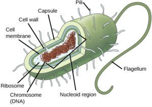
Unlike eukaryotes, bacteria have a cell wall made of peptidoglycan, comprised of sugars and amino acids, and many have a polysaccharide capsule (Figure 18.1). The cell wall acts as an extra layer of protection, helps the cell maintain its shape, and prevents dehydration. The capsule enables the cell to attach to surfaces in its environment. Some prokaryotes have flagella, pili, or fimbriae. Flagella are used for locomotion, while most pili are used to exchange genetic material during a type of reproduction called conjugation.

Components of Eukaryotic Cells

In nature, the relationship between form and function is apparent at all levels, including the level of the cell, and this will become clear as we explore eukaryotic cells. The principle “form follows function” is found in many contexts. For example, birds and fish have streamlined bodies that allow them to move quickly through the medium in which they live, be it air or water. It means that, in general, one can deduce the function of a structure by looking at its form, because the two are matched.

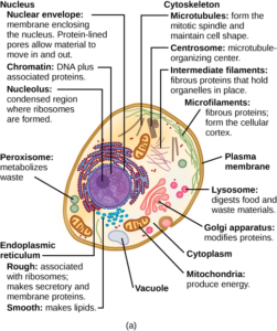
A eukaryotic cell is a cell that has a membrane-bound nucleus and other membrane-bound compartments or sacs, called organelles, which have specialized functions (Figures 18.2 and 18.3). The word eukaryotic means “true kernel” or “true nucleus,” pointing to the presence of the membrane-bound nucleus in these cells. The word “organelle” means “little organ,” and, as already mentioned, organelles have specialized cellular functions, just as the organs of your body have specialized functions. 

A cartoon diagram of an animal cell.
Figure 18.2. A typical animal cell.


Diagram of a typical plant cell.
Figure 18.3. A typical plant cell.

Video Transcript

On Earth, there are two major types of cells: eukaryote and prokaryote. Eukaryote cells are more complex in structure and function than prokaryote cells. In this tutorial, we’ll tour the major features of a eukaryotic cell using both an animal cell and a plant cell as examples.
Because of its large size, the nucleus is one of the most noticeable structures in the eukaryotic cell. The nucleus is the storage site for most of the cell’s genetic information. The dark structure inside the nucleus is the nucleolus. This is where the components of the ribosomes are manufactured.
The endoplasmic reticulum, or ER, is a membranous network of sac like structures in the cytoplasm of the cell. The ER is divided into two distinct types based upon structure and function.
The rough ER is studded with ribosomes, the location of protein synthesis in a cell. The presence of the ribosomes gives this ER its “rough” appearance. The proteins produced by the rough ER are often secreted by the cell and carried by vesicles to the Golgi apparatus for further modification.
The smooth ER is a membrane bound organelle involved in lipid synthesis, detoxification, and carbohydrate metabolism. It is given the name “smooth” endoplasmic reticulum because there are no ribosomes on its cytoplasmic surface.
The Golgi apparatus consists of a series of flattened sacs that resemble pancakes. Its job is to package and distribute cellular products that are destined for a specific location within the cell or for secretion. You can think of the Golgi apparatus as the “post office” of the cell, where packages are dropped off, and destinations are assigned.
Lysosomes, which are produced by the Golgi apparatus, are vesicles that aid in the breakdown of materials within the cell. They may also digest worn or damaged organelles and recycle their components.
Mitochondria are the location where the stored energy found in carbohydrates is used to produce cellular energy in the form of ATP. Mitochondria are found in both plant and animal cells.
Unique to the plant cells are the chloroplasts, the location of photosynthesis. This is where incoming sunlight is used to convert carbon dioxide gas to carbohydrates and other compounds.
Vacuoles are specialized membrane bound structures found mostly in the cells of plants. Vacuoles usually store substances, such as water, sugars and salts, as well as pigments and toxic molecules. The pigments are responsible for the red, blue and green colors of flowers and some leaves. The toxic chemicals keep the plant from being eaten by animals.
Centrioles are organelles found mainly in animal cells. They are composed of microtubules and occur in pairs. Centrioles function in the reorganization of microtubules during the process of cell division.
The cytoplasm contains a complex network of protein fibers called the cytoskeleton. These fibers include microtubules, microfilaments and intermediate filaments. Overall, the cytoskeleton plays a similar role to the bones and muscles in your body, providing structural support, and, along with motor proteins, allowing the cell and organelles to move.
Let’s do a quick recap. The internal structure of a eukaryotic cell is fairly complex and differs slightly between a plant cell and an animal cell. This diagram will help you remember some of the similarities and differences.

Cell Size

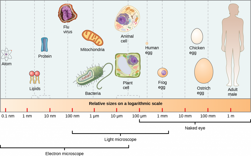
At 0.1–5.0 μm in diameter, prokaryotic cells are significantly smaller than eukaryotic cells, which have diameters ranging from 10–100 μm (Figure 18.4). The small size of prokaryotes allows ions and organic molecules that enter them to quickly spread to other parts of the cell. Similarly, any wastes produced within a prokaryotic cell can quickly move out. However, larger eukaryotic cells have evolved different structural adaptations to enhance cellular transport. Indeed, the large size of these cells would not be possible without these adaptations. In general, cell size is limited because volume increases much more quickly than does cell surface area. As a cell becomes larger, it becomes more and more difficult for the cell to acquire sufficient materials to support the processes inside the cell, because the relative size of the surface area across which materials must be transported declines.

infographic showing sizes of structures at different scales
Figure 18.4. Relative sizes of various structures.
Figure 18.4 Image Description

Across the bottom are different size labels from 0.1nm to 1m on the right. Above this is a label “relative sizes on a logarithmic scale. Farthest left over 0.1nm is an illustration of an atom. Above 1nm are phospholipids shown as red circles with two yellow lines extending down. Proteins are above 10nm. A purple blob labeled flu virus is located above 100nm. An oval-shaped mitochondrion and a green oval-shaped bacteria are above 1 micrometer. Diagrams of animal and plant cells are between 10 and 100 micrometers. A purple circle inside a peach circle is labeled frog egg and is above 1mm. A white egg shape labeled chicken egg is between 10mm and 100mm. A diagram of an adult human is above 1m.

References

Unless otherwise noted, images on this page are licensed under CC-BY 4.0 by OpenStax.

Text adapted from: OpenStax, Concepts of Biology. OpenStax CNX. May 18, 2016. http://cnx.org/contents/b3c1e1d2-839c-42b0-a314-e119a8aafbdd@9.10

License

Icon for the Creative Commons Attribution 4.0 International License

MHCC Biology 112: Biology for Health Professions Copyright © 2019 by Lisa Bartee is licensed under a Creative Commons Attribution 4.0 International License, except where otherwise noted.

Share This Book