
15 Nucleic Acids
Nucleic acids are key macromolecules in the continuity of life. They carry the genetic blueprint of a cell and carry instructions for the functioning of the cell. The two main types of nucleic acids are deoxyribonucleic acid (DNA) and ribonucleic acid (RNA). DNA is the genetic material found in all living organisms, ranging from single-celled bacteria to multicellular mammals. The other type of nucleic acid, RNA, is mostly involved in protein synthesis. The DNA molecules never leave the nucleus, but instead use an RNA intermediary to communicate with the rest of the cell. Other types of RNA are also involved in protein synthesis and its regulation. We will be going into more detail about nucleic acids in a later section.
DNA and RNA are made up of monomers known as nucleotides connected together in a chain with covalent bonds. Each nucleotide is made up of three components: a nitrogenous base, five-carbon sugar, and a phosphate group (Figure 1). The nitrogenous base in one nucleotide is attached to the sugar molecule, which is attached to the phosphate group.
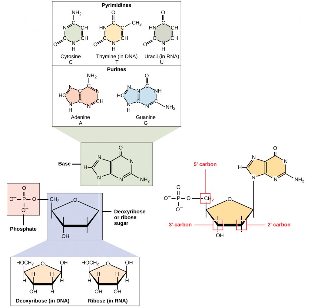
The nitrogenous bases, important components of nucleotides, are organic molecules and are so named because they contain carbon and nitrogen. They are bases because they contain an amino group that has the potential of binding an extra hydrogen, and thus, decreases the hydrogen ion concentration in its environment, making it more basic. Each nucleotide in DNA contains one of four possible nitrogenous bases: adenine (A), guanine (G) cytosine (C), and thymine (T). RNA contains the base uracil (U) instead of thymine. The order of the bases in a nucleic acid determines the information that the molecule of DNA or RNA carries. This is because the order of the bases in a DNA gene determines the order that amino acids will be assembled together to form a protein.
The pentose sugar in DNA is deoxyribose, and in RNA, the sugar is ribose (Figure 1). The difference between the sugars is the presence of the hydroxyl group on the second carbon of the ribose and hydrogen on the second carbon of the deoxyribose. The carbon atoms of the sugar molecule are numbered as 1′, 2′, 3′, 4′, and 5′ (1′ is read as “one prime”). The phosphate residue is attached to the hydroxyl group of the 5′ carbon of one sugar and the hydroxyl group of the 3′ carbon of the sugar of the next nucleotide, which forms a 5′–3′ phosphodiester linkage (a specific type of covalent bond). A polynucleotide may have thousands of such phosphodiester linkages.
DNA Double-Helical Structure
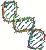
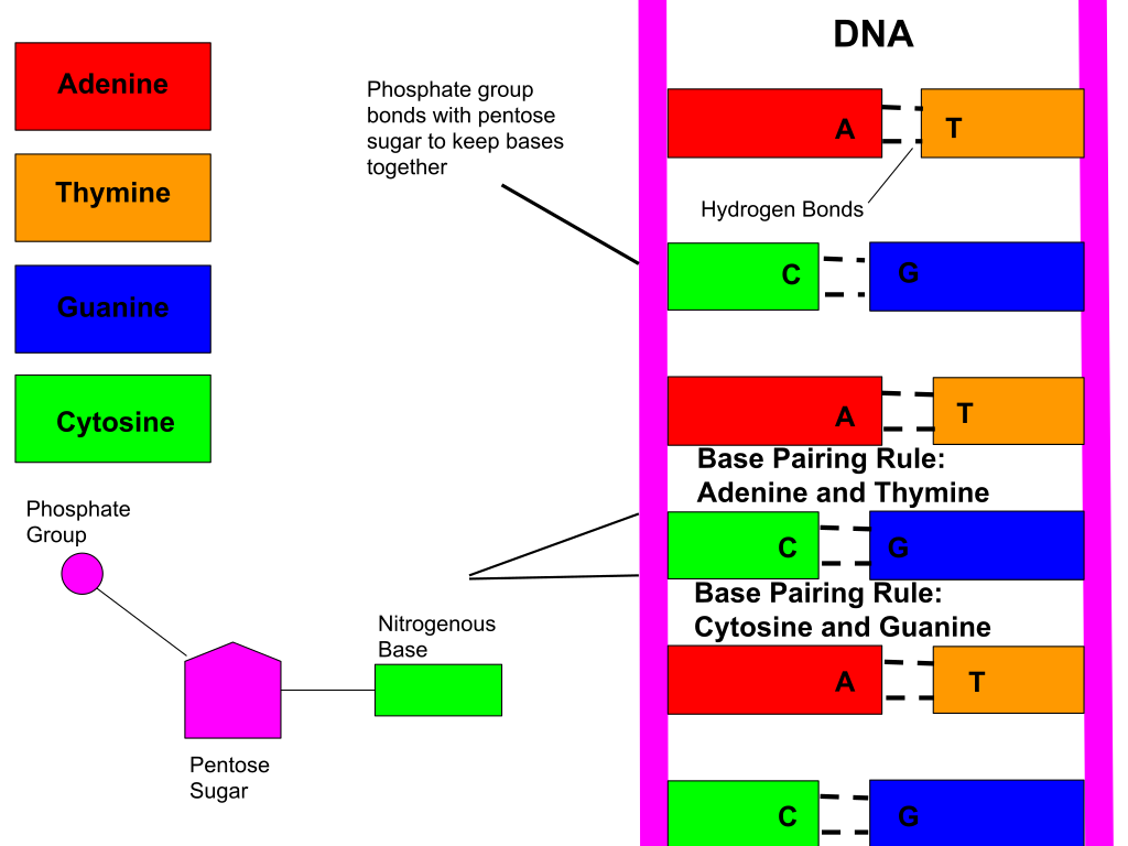
DNA has a double-helical structure (Figure 2). It is composed of two strands, or chains, of nucleotides. The double helix of DNA is often compared to a twisted ladder. The strands (the outside parts of the ladder) are formed by linking the phosphates and sugars of adjacent nucleotides with strong chemical bonds, called covalent bonds. The rungs of the twisted ladder are made up of the two bases attached together with a weak chemical bond, called a hydrogen bonds. Two bases hydrogen bonded together is called a base pair. The ladder twists along its length, hence the “double helix” description, which means a double spiral.
The alternating sugar and phosphate groups lie on the outside of each strand, forming the backbone of the DNA. The nitrogenous bases are stacked in the interior, like the steps of a staircase, and these bases pair; the pairs are bound to each other by hydrogen bonds. The bases pair in such a way that the distance between the backbones of the two strands is the same all along the molecule.
In a molecule of DNA, adenine (A) always pairs with thymine (T), and cytosine (C) always pairs with guanine (G). This means that the sequence of one strand of the DNA double helix can always be used to determine the other strand.
How does nucleic acid structure determine function?
The major function of both DNA and RNA is to store and carry genetic information. The specific order of nucleotides in the molecule of DNA or RNA is what determines the genetic information it carries. You can think of it like letters in a book – if the order of the letters were changed, the book would no longer contain the same (or correct) information.
References
Unless otherwise noted, images on this page are licensed under CC-BY 4.0 by OpenStax.
OpenStax, Biology. OpenStax CNX. May 27, 2016. https://cnx.org/contents/GFy_h8cu@10.120:U7tPDRxK@9/DNA-Structure-and-Sequencing
