"

83 Meiosis II

In some species, cells enter a brief interphase, or interkinesis, before entering meiosis II. Interkinesis lacks an S phase, so chromosomes are not duplicated. The two cells produced in meiosis I go through the events of meiosis II at the same time. During meiosis II, the sister chromatids within the two daughter cells separate, forming four new haploid gametes, each with one copy of each chromosome. The mechanics of meiosis II is similar to mitosis, except that each dividing cell has only one set of homologous chromosomes. Therefore, each cell has half the number of sister chromatids to separate out as a diploid cell undergoing mitosis.

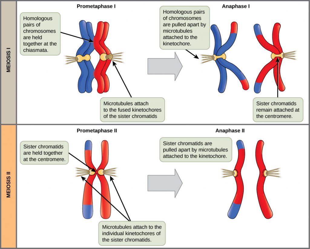
During meiosis II, each sister chromatid is attached to spindle fiber microtubules from opposite poles. The sister chromatids are pulled apart by the spindle fiber microtubules and move toward opposite poles (Figure 83.1). 


diagram differentiating meiosis I from meiosis II
Figure 83.1. In prometaphase I, microtubules attach to the fused kinetochores of homologous chromosomes. In anaphase I, the homologous chromosomes are separated. In prometaphase II, microtubules attach to individual kinetochores of sister chromatids. In anaphase II, the sister chromatids are separated.
Figure 83.1 Image Description

The diagram illustrates prometaphase I and II and anaphase I and II, which occur in Meiosis I and Meiosis II. The top panel illustrates Prometaphase I and Anaphase I in Meiosis I. In Prometaphase I, a red and a blue set of homologous chromosomes (each made up of 2 sister chromatids) are shown with yellow microtubules attached to their centromeres. The homologous pairs of chromosomes are held together at the chiasmata, which are the regions where crossing over will occur. Microtubules attach to the fused kinetochores of the sister chromatids. To the right of this is Anaphase I, when homologous pairs of chromosomes are pulled apart by microtubules attached to the kinetochore. The two sister chromatids making up each chromosome remain attached at the centromere, but now one of the red chromatids has a blue tip at either end and one of the blue chromatids has a red tip at either end. This illustrates that crossing over has taken place, which has swapped sections of the blue and red chromosomes.

The lower panel illustrates Prometaphase II and Anaphase I Iin Meiosis II. In Prometaphase II, only the red homologous chromosome (with blue tips on one chromatid) is shown. The sister chromatids are still held together at the centromere. Microtubules are attached to the individual kinetochores of the sister chromatids. In Anaphase II, the sister chromatids are pulled apart by the microtubules attached to the kinetochore. The two sister chromatids are not identical because one has blue tips from crossing over with the blue homologous chromosome.

The chromosomes arrive at opposite ends of the cells and begin to decondense (unwind). Nuclear envelopes form around the chromosomes. Cytokinesis separates the two cells into four unique haploid cells. At this point, the newly formed nuclei are both haploid and have only one copy of the single set of chromosomes. The cells produced are genetically unique because of the random assortment of paternal and maternal homologs and because of the recombining of maternal and paternal segments of chromosomes (with their sets of genes) that occurs during crossover.

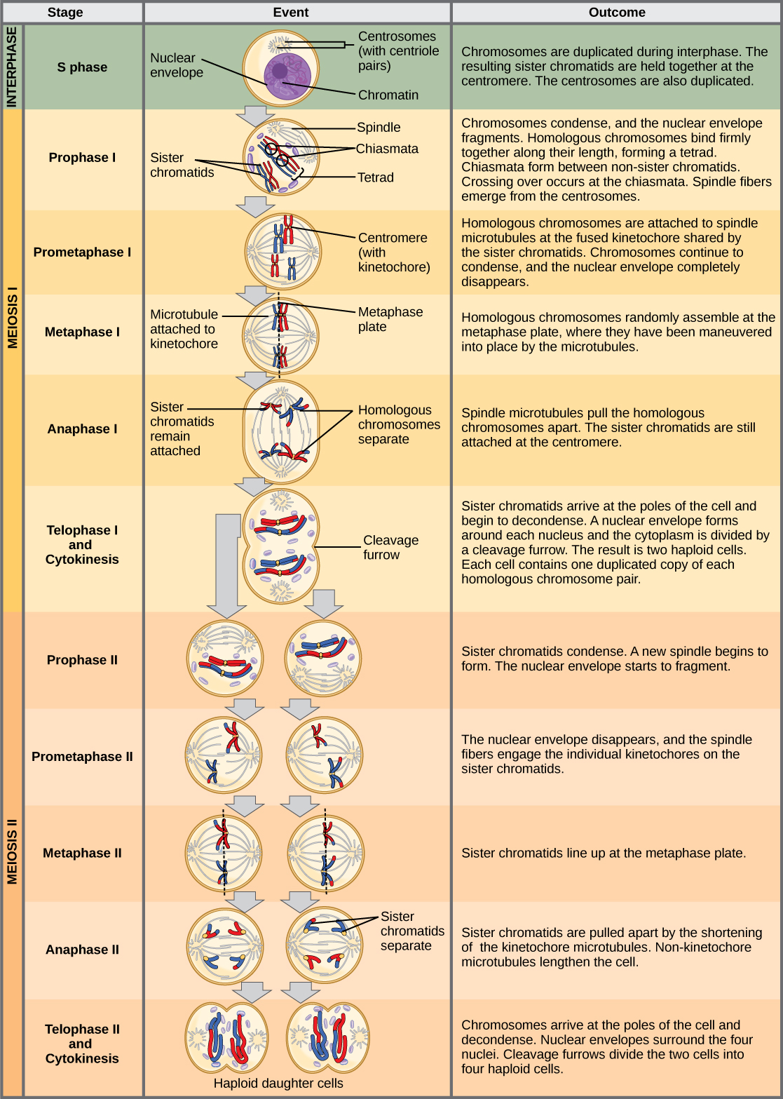
The entire process of meiosis is outlined in Figure 83.2 (you do not need to know the names of the phases or what happens during each phase, only what happens overall during meiosis I and II).

meiosis
Figure 83.2. An animal cell with a diploid number of four (2n = 4) proceeds through the stages of meiosis to form four haploid daughter cells.
Figure 83.2 Image Description

This diagram summarizes the events that occur during each stage of meiosis. The top section of the diagram is green and is labeled Interphase; S phase. The cell has a nucleus surrounded by a nuclear envelope and the chromatin is evenly distributed throughout the nucleus. The outcome of S phase is that chromosomes are duplicated during interphase. The resulting sister chromatids are held together at the centromere. The centrosomes are also duplicated. The next section of the diagram is yellow and has several stages identified. In Prophase I, chromosomes condense and the nuclear envelope fragments. Homologous chromosomes bind firmly together along their length, forming a tetrad. Chiasmata form between non-sister chromatids. Crossing over occurs at the chiasmata. Spindle fibers emerge from the centrosomes. In Prometaphase I, Homologous chromosomes are attached to spindle microtubules at the fused kinetochore shared by the sister chromatids. Chromosomes continue to condense, and the nuclear envelope completely disappears. In Metaphase I, homologous chromosomes randomly assemble at the metaphase plate, where they have been maneuvered into place by the microtubules.

In Anaphase I, the sister chromatids remain attached together at the centromeres, while the homologous chromosomes are pulled apart towards opposite ends of the cell by spindle microtubules. In Telophase I and Cytokinesis, the sister chromatids arrive at the poles of the cell and begin to decondense. A nuclear envelope forms around each nucleus and the cytoplasm is divided by a cleavage furrow. The result is two haploid cells. Each cell contains one duplicated copy of each homologous chromosome pair. Meiosis II begins with Prophase II, which occurs in both of the daughter cells produced at the end of telophase I and cytokinesis. The sister chromatids condense and a new spindle begins to form. The nuclear envelope starts to fragment. In Prometaphase II, the nuclear envelope disappears and the spindle fibers engage the individual kinetochores on the sister chromatids. In Metaphase II, the sister chromatids line up at the metaphase plate. During Anaphase II, the sister chromatids are pulled apart by the shortening of the kinetochore microtubules. Non-kinetochore microtubules lengthen the cell. In Telophase II and Cytokinesis, the chromosomes arrive at the poles of the cell and decondense. Nuclear envelopes surround the four nuclei. Cleavage furrows divide the two cells into four haploid cells.

Summary of Meiosis II

Meiosis II begins with the 2 haploid cells where each chromosome is made up of two connected sister chromatids. DNA replication does NOT occur at the beginning of meiosis II. The sister chromatids are separated, producing 4 genetically different haploid cells.

References

Unless otherwise noted, images on this page are licensed under CC-BY 4.0 by OpenStax.

OpenStax, Biology. OpenStax CNX. May 27, 2016. http://cnx.org/contents/s8Hh0oOc@9.10:1Q8z96mT@4/Meiosis

License

Icon for the Creative Commons Attribution 4.0 International License

MHCC Biology 112: Biology for Health Professions Copyright © 2019 by Lisa Bartee is licensed under a Creative Commons Attribution 4.0 International License, except where otherwise noted.

Share This Book