
Protein Structure
What Is Protein?
Proteins are macromolecules composed of amino acids. For this reason, amino acids are commonly called the building blocks of protein. There are 20 different amino acids, and we require all of them to make the many different proteins found throughout the body. Proteins are crucial for the nourishment, renewal, and continuance of life.
Just like carbohydrates and fats, proteins contain the elements carbon, hydrogen, and oxygen, but proteins are the only macronutrient that also contain nitrogen as part of their core structure. In each amino acid, the elements are arranged into a specific conformation, consisting of a central carbon bound to the following four components:
- A hydrogen
- A nitrogen-containing amino group
- A carboxylic acid group (hence the name “amino acid”)
- A side chain
The first three of those components are the same for all amino acids. The side chain—represented by an “R” in the diagram below—is what makes each amino acid unique.
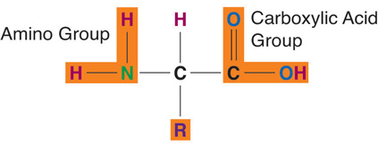
Figure 6.1. Amino Acid Structure.
Amino acid side chains vary tremendously in their size and can be as simple as one hydrogen (as in glycine, shown in Figure 6.1) or as complex as multiple carbon rings (as in tryptophan). They also differ in their chemical properties, thus impacting the way amino acids act in their environment and with other molecules. Because of their side chains, some amino acids are polar, making them hydrophilic and water-soluble, whereas others are nonpolar, making them hydrophobic or water-repelling. Some amino acids carry a negative charge and are acidic, while others carry a positive charge and are basic. Some carry no charge. Some examples are given below. For this class, you don’t need to memorize amino acid structures or names, but you should appreciate the diversity of amino acids and understand that it is the side chain that makes each different.
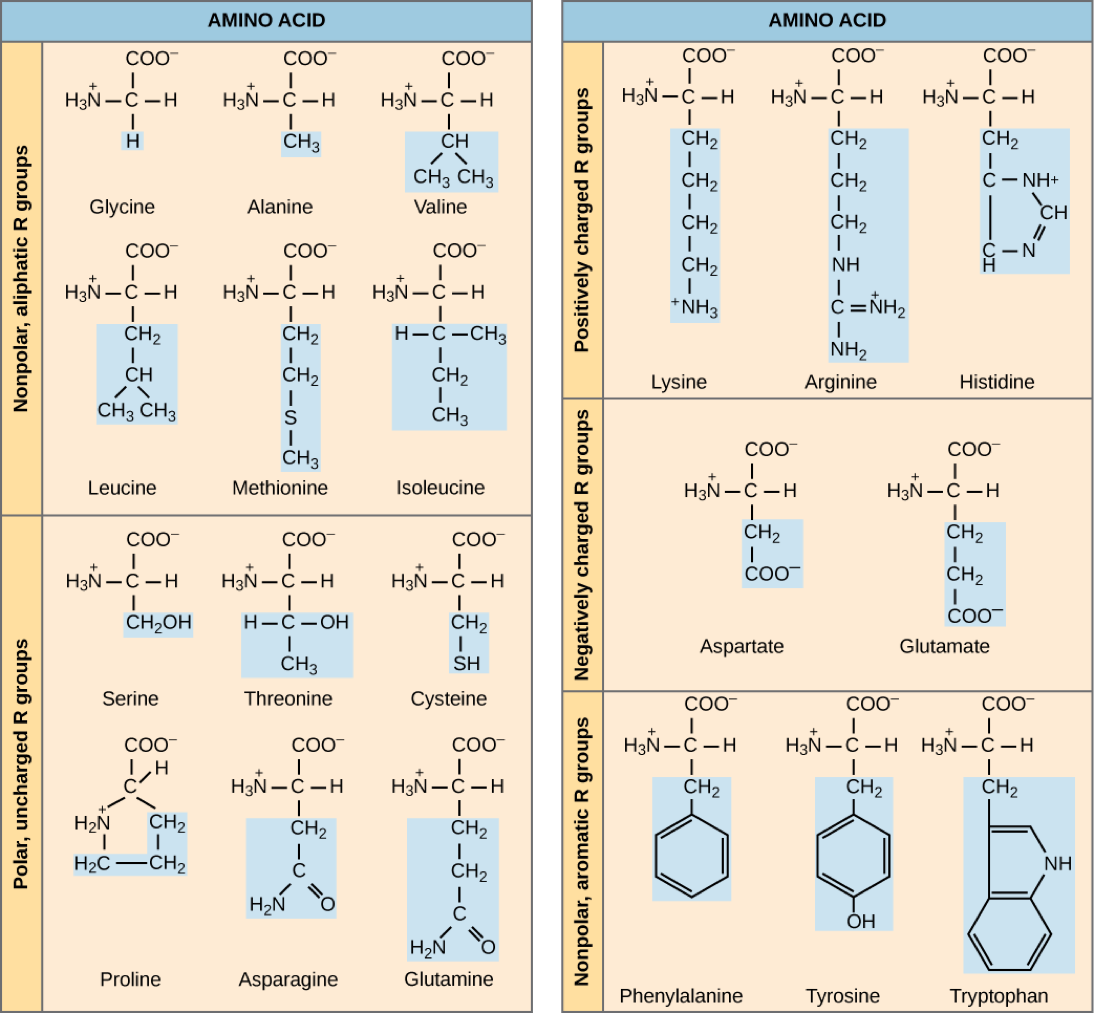
Figure 6.2. Amino acids have different structures and chemical properties, determined by their side chains.
Essential and Nonessential Amino Acids
We also classify amino acids based on their nutritional aspects (Table 6.1 “Essential and Nonessential Amino Acids”):
- Nonessential amino acids are not required in the diet, because the body can synthesize them. They’re still vital to protein synthesis, and they’re still present in food, but because the body can make them, we don’t have to worry about nutritional requirements. There are 11 nonessential amino acids.
- Essential amino acids can’t be synthesized by the body in sufficient amounts, so they must be obtained in the diet. There are 9 essential amino acids.
|
Essential |
Nonessential |
|
Histidine |
Alanine |
|
Isoleucine |
Arginine* |
|
Leucine |
Asparagine |
|
Lysine |
Aspartic Acid |
|
Methionine |
Cysteine* |
|
Phenylalanine |
Glutamic Acid |
|
Threonine |
Glutamine |
|
Tryptophan |
Glycine* |
|
Valine |
Proline* |
|
|
Serine |
|
|
Tyrosine* |
|
*Conditionally essential |
|
Table 6.1. Essential and nonessential amino acids
Sometimes during infancy, growth, and in diseased states, the body cannot synthesize enough of some of the nonessential amino acids and more of them are required in the diet. These types of amino acids are called conditionally essential amino acids.
The nutritional value of a protein is dependent on what amino acids it contains and in what quantities. As we’ll discuss later, a food that contains all of the essential amino acids in adequate amounts is called a complete protein source, whereas one that does not is called an incomplete protein source.
The Many Different Types of Proteins
There are over 100,000 different proteins in the human body. Proteins are similar to carbohydrates and lipids in that they are polymers (simple repeating units); however, proteins are much more structurally complex. In contrast to carbohydrates, which have identical repeating units, proteins are made up of amino acids that are different from one another. Different proteins are produced because there are 20 types of naturally occurring amino acids that are combined in unique sequences.
Additionally, proteins come in many different sizes. The hormone insulin, which regulates blood glucose, is composed of only 51 amino acids. On the other hand, collagen, a protein that acts like glue between cells, consists of more than 1,000 amino acids. Titin is the largest known protein. It accounts for the elasticity of muscles and consists of more than 25,000 amino acids!
The huge diversity of proteins is also due to the unending number of amino acid sequences that can be formed. To understand how so many different proteins can be made from only 20 amino acids, think about music. All of the music that exists in the world has been derived from a basic set of seven notes C, D, E, F, G, A, B (with the addition of sharps and flats), and there is a vast array of music all composed of specific sequences from these basic musical notes. Similarly, the 20 amino acids can be linked together in an extraordinary number of sequences. For example, if an amino acid sequence for a protein is 104 amino acids long, the possible combinations of amino acid sequences is equal to 20104, which is 2 followed by 135 zeros!
Building Proteins with Amino Acids
The decoding of genetic information to synthesize a protein is the central foundation of modern biology. The building of a protein consists of a complex series of chemical reactions that can be summarized into three basic steps: transcription, translation, and protein folding.
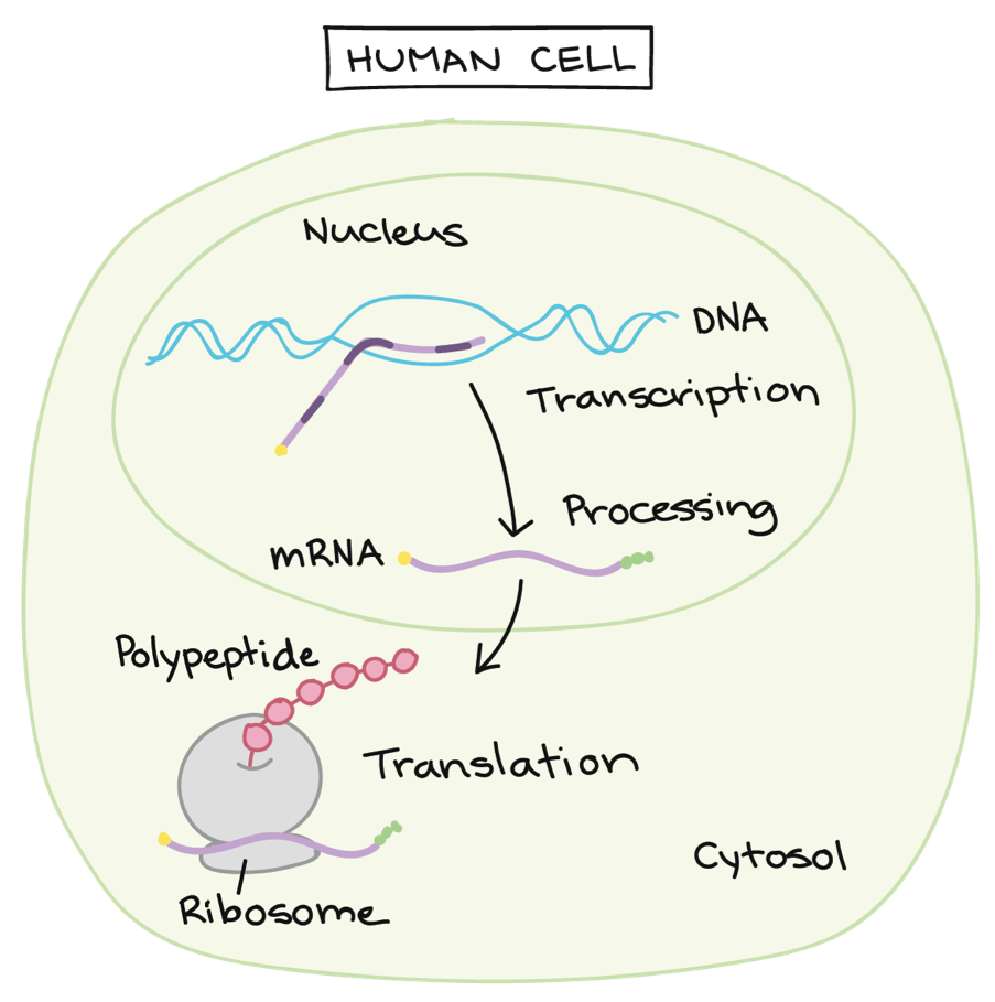
Figure 6.3. Overview of protein synthesis. Protein folding happens after translation.
- Transcription – Deoxyribonucleic acid, or DNA, is the long, double-stranded molecules containing your genome—instructions for making all of the proteins in your body. In the nucleus of the cell, the DNA must be transcribed or copied into the single-stranded messenger ribonucleic acid (mRNA), which carries the genetic instructions into the cell’s cytosol for protein synthesis.
- Translation – At the ribosomes in the cell’s cytosol, amino acids are linked together in the specific order dictated by the mRNA. Each amino acid is connected to the next amino acid by a special chemical bond called a peptide bond(Figure 6.4). The peptide bond forms between the carboxylic acid group of one amino acid and the amino group of another, releasing a molecule of water. As amino acids are linked sequentially by peptide bonds, following the specific order dictated by the mRNA, the protein chain, also known as a polypeptide chain, is built (Figure 6.5).
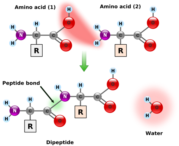
Figure 6.4. Peptide bond formation
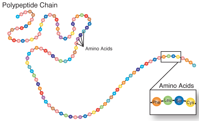
Figure 6.5. A polypeptide chain
3. Protein folding – The polypeptide chain folds into specific three-dimensional shapes, as described in the next section.
VIDEO: “DNA Transcription,” by DNA Learning Center, YouTube (March 22, 2010), 1:52 minutes.
VIDEO: “mRNA Translation,” DNA Learning Center, YouTube (March 22, 2010), 2:04 minutes.
Protein Organization
Protein’s structure enables it to perform a variety of functions. There are four different structural levels of proteins (Figure 6.6.):
- Primary structure – This is the one-dimensional polypeptide chain of amino acids, held together by peptide bonds.
- Secondary structure – The polypeptide chain folds into simple coils (also called helices) and sheets, determined by the chemical interactions between amino acids.
- Tertiary structure – This is the unique three-dimensional shape of a protein, formed as the different side chains of amino acids chemically interact, either repelling or attracting each other. Thus, the sequence of amino acids in a protein directs the protein to fold into a specific, organized shape.
- Quaternary structure – In some proteins, multiple folded polypeptides called subunits combine to make one larger functional protein. This is called quaternary protein structure. The protein hemoglobin is an example of a protein that has quaternary structure. It is composed of four polypeptides that bond together to form a functional oxygen carrier.
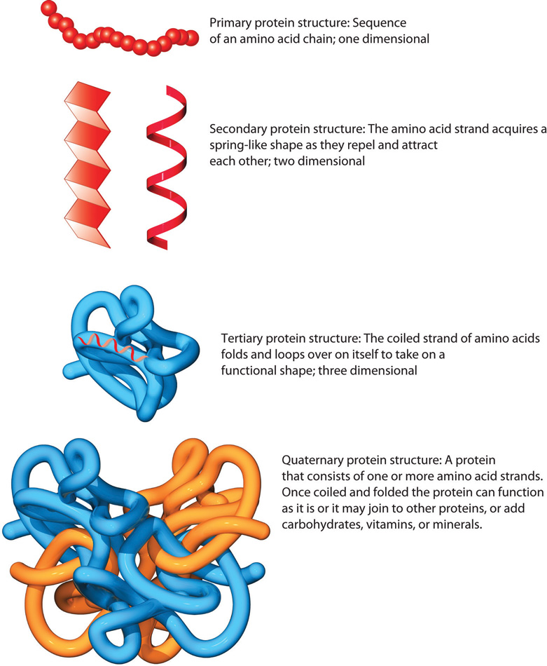
Figure 6.6. A protein has four different structural levels.
VIDEO: “What is a Protein,” by RCSBProteinDataBank, YouTube (September 4, 2016), 2:38 minutes. This video gives an overview of the structure of amino acids, the four different structural levels of protein, and examples of different types of proteins in the body.
A protein’s structure also influences its nutritional quality. Large fibrous protein structures are more difficult to digest than smaller proteins and some, such as keratin, are indigestible. Because digestion of some fibrous proteins is incomplete, not all of the amino acids are absorbed and available for the body to utilize, thereby decreasing their nutritional value.
The specific three-dimensional structure of proteins can be disrupted by changes in their physical environment, causing them to unfold. This is called denaturation, and it results in loss of both structure and function of proteins. Changes in pH (acidic or basic conditions) and exposure to heavy metals, alcohol, and heat can all cause protein denaturation. The proteins in cooked foods are at least partially denatured from the heat of cooking, and denaturation in the stomach is an important part of protein digestion, as we’ll discuss later in this unit. We can see everyday examples of denaturation in cooking techniques, like how egg whites become solid and opaque with cooking, and cream becomes fluffy when it’s whipped. Both of these are examples of denaturation leading to physical changes in protein structure, and because protein structure determines function, denaturation also causes proteins to lose their function.
Shape Determines Function
An important concept with proteins is that SHAPE determines FUNCTION. A change in the amino acid sequence will cause a change in protein shape. Each protein in the human body differs in its amino acid sequence and consequently, its shape. The synthesized protein is structured to perform a particular function in a cell. A protein made with an incorrectly placed amino acid may not function properly, and this can sometimes cause disease. An example of this is sickle cell anemia, a genetic disorder. Below is a picture of hemoglobin, a protein with a globular three-dimensional structure. When packed in red blood cells to deliver oxygen, this structure gives red blood cells a donut shape.
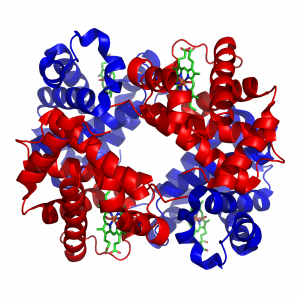
Figure 6.7. Structure of hemoglobin
In people with sickle cell anemia, DNA gives cells the incorrect message when bonding amino acids together to make hemoglobin. The result is crescent-shaped red blood cells that are sticky and do not transport oxygen like normal red blood cells, as illustrated in the figure below.
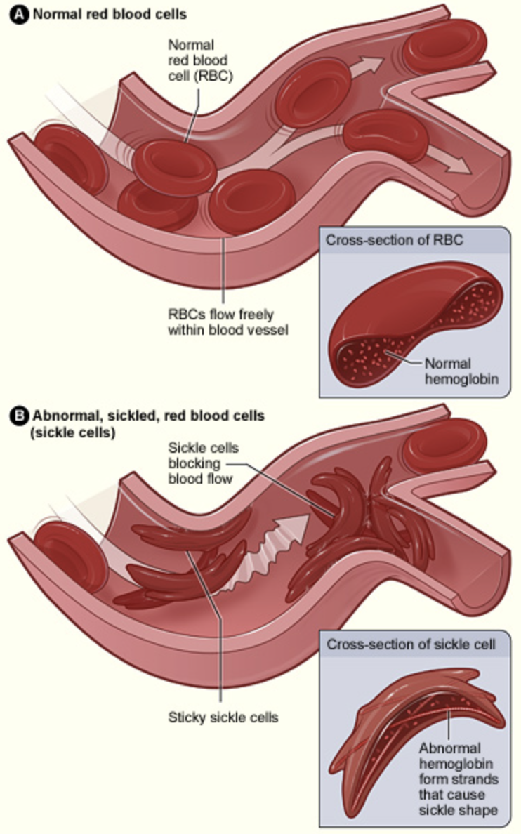
Figure 6.8. Difference in blood cells and blood flow between normal red blood cells and sickle shaped blood cells.
Self-Check
Attributions:
- Lindshield, B. L. Kansas State University Human Nutrition (FNDH 400) Flexbook. goo.gl/vOAnR, CC BY-NC-SA 4.0
- “Defining Protein,” section 6.1 from the book An Introduction to Nutrition (v. 1.0), CC BY-NC-SA 3.0
Image Credits:
- Figure 6.1. “Amino acid structure” from “Defining Protein”, section 6.1 from the book An Introduction to Nutrition (v. 1.0) is licensed under CC BY-NC-SA 3.0
- Figure 6.2. “Amino acids diagram” from Section 3.4 of Biology by OpenStax is licensed under CC BY 4.0
- Table 6.1. “Essential and nonessential amino acids” by Tamberly Powell is licensed under CC BY-NC-SA 2.0
- Figure 6.3. “Protein synthesis diagram” from “Intro to gene expression (central dogma)” by Khan Academy is licensed under CC BY-NC-SA 4.0
- Figure 6.4. “Polypeptide chain” by the NIH is in the Public Domain
- Figure 6.5. “Peptide bond formation” by Yassine Mrabet is in the Public Domain
- Figure 6.6. “Structural levels of protein” from “Defining Protein”, section 6.1 from the book An Introduction to Nutrition (v. 1.0) is licensed under CC BY-NC-SA 3.0
- Figure 6.7. “Hemoglobin” by Richard Wheeler is licensed under CC BY-SA 3.0
- Figure 6.8. “Blood flow and red blood cell shape of normal and sickle cell shaped hemoglobin” by The National Heart, Lung, and Blood Institute (NHLBI) is in the Public Domain
Macromolecules composed of chains of amino acids, which are simple subunits made of carbon, oxygen, hydrogen, and nitrogen.
The building blocks of protein.
Amino acids that are not required in the diet because the body can synthesize them.
Amino acids that must be obtained in the diet because they can’t be synthesized by the body in sufficient amounts.
Amino acids that must be obtained in the diet in certain situations when more are needed than the body can synthesize.
A food that contains all of the essential amino acids in adequate amounts needed by the body.
A food that does not contain all of the essential amino acids in adequate amounts needed by the body.
The first major step of protein synthesis; a process in which the genetic code of DNA is copied into messenger RNA.
The second major step of protein synthesis; information on messenger RNA is translated into building a protein.
A special chemical bond between two amino acids.
The third major step of protein synthesis; the amino acid chain folds into three-dimensional shapes.
One-dimensional polypeptide chain of amino acids.
Polypeptide chain that has folded into two-dimensional simple coils (also called helices) and sheets.
Polypeptide chain that has folded into a three-dimensional organized shape based on the interactions of the amino acids.
Multiple folded polypeptides called subunits that have combined to make one larger functional protein.
When the three-dimensional structure of a protein is unfolded due to a change in the environment (e.g., acid, heat); results in loss of protein function.
