
The Cytoskeleton
If you were to remove all the organelles from a cell, would the plasma membrane and the cytoplasm be the only components left? No. Within the cytoplasm, there would still be ions and organic molecules, plus a network of protein fibers known as the cytoskeleton.
Both prokaryotes and eukaryotes have a cytoskeleton. Both types of organisms use their cytoskeleton for cell division, protection, and shape determination.
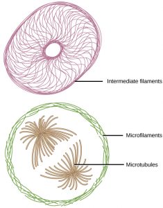
In addition, in eukaryotes the cytoskeleton also functions to secure certain organelles in specific positions, and to allow cytoplasm and vesicles to move within the cell. It also enables unicellular organisms to move independently. There are three types of fibers within the cytoskeleton: microfilaments, also known as actin filaments, intermediate filaments, and microtubules (Figure 1).
Microfilaments
Of the three types of protein fibers in the cytoskeleton, microfilaments are the narrowest. They function in cellular movement, have a diameter of about 7 nm, and are made of two intertwined strands of a globular protein called actin. For this reason, microfilaments are also known as actin filaments.
ATP is required for actin proteins to assemble into long filaments. These long actin filaments serve as a track for the movement of a motor protein called myosin. Actin and myosin are plentiful in muscle cells. When your actin and myosin filaments slide past each other, your muscles contract. Actin also enables your cells to engage in cellular events requiring motion, such as cell division in animal cells and cytoplasmic streaming, which is the circular movement of the cell cytoplasm in plant cells.
Microfilaments also provide some rigidity and shape to the cell. They can depolymerize (disassemble) and reform quickly, thus enabling a cell to change its shape and move. White blood cells (your body’s infection-fighting cells) make good use of this ability. They can move to the site of an infection and phagocytize the pathogen.
Intermediate Filaments
Intermediate filaments are made of several strands of fibrous proteins that are wound together. These elements of the cytoskeleton get their name from the fact that their diameter, 8 to 10 nm, is between those of microfilaments and microtubules.
Intermediate filaments have no role in cell movement. Their function is purely structural. They bear tension, thus maintaining the shape of the cell, and anchor the nucleus and other organelles in place. Figure 1 shows how intermediate filaments create a supportive scaffolding inside the cell.
The intermediate filaments are the most diverse group of cytoskeletal elements. Several types of fibrous proteins are found in the intermediate filaments. You are probably most familiar with keratin, the fibrous protein that strengthens your hair, nails, and the epidermis of the skin.
Microtubules
As their name implies, microtubules are small hollow tubes. The walls of the microtubule are made of polymerized dimers of α-tubulin and β-tubulin, two globular proteins. With a diameter of about 25 nm, microtubules are the widest components of the cytoskeleton. They help the cell resist compression, provide a track along which vesicles move through the cell, and pull replicated chromosomes to opposite ends of a dividing cell. Like microfilaments, microtubules can dissolve and reform quickly.
Microtubules are also the structural elements of flagella, cilia, and centrioles (the latter are the two perpendicular bodies of the centrosome). In fact, in animal cells, the centrosome is the microtubule-organizing center. In eukaryotic cells, flagella and cilia are quite different structurally from their counterparts in prokaryotes, as discussed below.
The centrosome replicates itself before a cell divides, and the centrioles play a role in pulling the duplicated chromosomes to opposite ends of the dividing cell. However, the exact function of the centrioles in cell division is not clear, since cells that have the centrioles removed can still divide, and plant cells, which lack centrioles, are capable of cell division.
References
Unless otherwise noted, images on this page are licensed under CC-BY 4.0 by OpenStax.
Text adapted from: OpenStax, Concepts of Biology. OpenStax CNX. May 18, 2016 http://cnx.org/contents/b3c1e1d2-839c-42b0-a314-e119a8aafbdd@9.10
